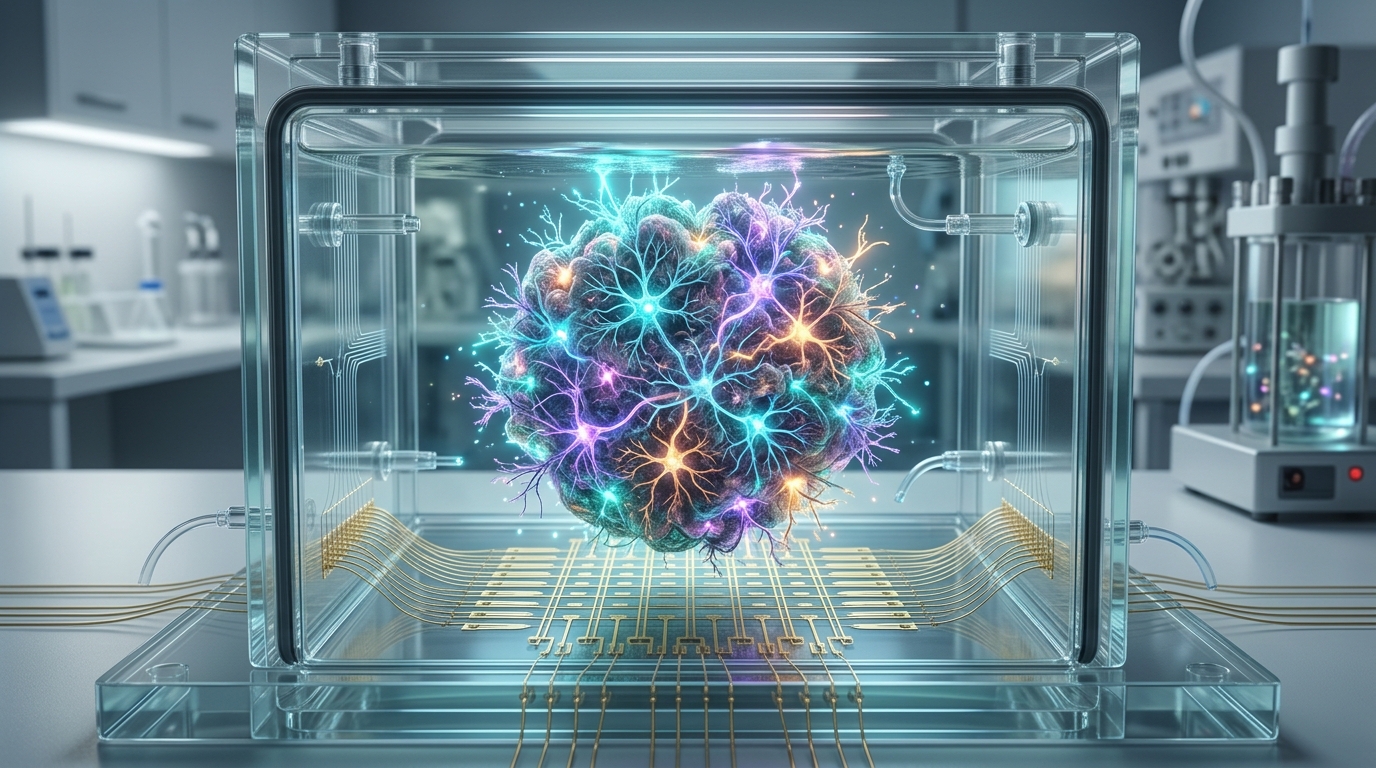
Human neural organoids, the three-dimensional clusters of stem-cell-derived neurons that self-organize into brain-like tissue, have moved beyond static models of development into systems whose electrical activity can be directly measured. A new review in Nature Reviews Bioengineering synthesizes the technologies now used to interrogate these living networks, from calcium and voltage imaging to patch clamp electrophysiology and multielectrode array recordings, and argues that functional readouts are becoming the defining measure of how faithfully organoids reproduce human neural biology.
The appeal of organoids lies in what conventional systems cannot offer. Animal models and flat, two-dimensional cultures have informed neuroscience for decades, but they cannot reproduce the genetic, developmental and architectural complexity of the human brain. Organoids bridge that gap by allowing researchers to record neuronal activity at three distinct scales: single cells, local ensembles and whole networks. According to the review, bioengineering advances that improve organoid viability and electrode–tissue coupling have made this functional interrogation possible across all three levels.
The optical toolkit is described first. Calcium imaging exploits fluorescent dyes or genetically encoded indicators whose brightness shifts when neurons fire, while voltage imaging tracks membrane potential changes more directly and at faster timescales. These approaches generate large-scale activity maps across intact, three-dimensional tissue, revealing how signals propagate through developing circuits. Electrophysiological methods complement them: patch clamp recording measures the electrical behaviour of individual neurons with high precision, and multielectrode arrays, including next-generation architectures, capture network-wide firing patterns over days or weeks without damaging the tissue.
Assay selection, the authors emphasize, is not straightforward. The three-dimensional geometry of organoids, their cellular heterogeneity and the fragility of tissue handling all shape which measurements are feasible. Accessibility-enhancing formats such as tissue slicing and surface coupling improve signal quality by bringing sensors closer to active cells, but they can alter the microenvironment and disrupt connectivity. The review stresses that such trade-offs demand careful interpretation and transparent reporting, since a cleaner signal may come at the cost of an altered network.
Two strategies extend what organoids can do. Multi-regional assembloids, in which organoids representing different brain regions are fused, provide a context for studying inter-regional connectivity, the long-range interactions that define real brains. Xenotransplantation, in which human organoid tissue is grafted into animal hosts, promotes enhanced functional maturation and vascularization. The review positions both approaches as complementary platforms and as practical benchmarks for the limits of in vitro maturation, showing how far cultured tissue can be pushed toward adult-like function.
Perhaps the most consequential shift is computational. By integrating analytical frameworks with functional measurements, researchers can extract quantitative features such as excitability, synchrony and network topology from raw recordings. These metrics link molecular programs, the gene expression patterns that drive organoid development, to emergent circuit behaviour, turning descriptive observations into testable mechanistic hypotheses. The review argues that this quantitative layer is what elevates organoid studies from phenomenology to systems neuroscience.
Emerging findings push further still. The review highlights demonstrations of synaptic plasticity, the ability of connections to strengthen or weaken with activity, and of adaptive computation and goal-directed learning in closed-loop paradigms, where organoid networks receive feedback and adjust their output. Such experiments extend organoid research beyond characterizing spontaneous activity into territory once reserved for trained animal models. The authors caution, however, that these learning-like behaviours remain technically and conceptually nascent and should be interpreted conservatively, a restraint that distinguishes the review from more breathless coverage of in vitro learning claims.
Functional organoids also promise to reshape disease modelling. By revealing mechanisms of network dysfunction, they can illuminate neurodevelopmental disorders such as autism and epilepsy, neuropsychiatric conditions such as schizophrenia, and neurodegenerative diseases, all in a human, species-specific context that animal models approximate only imperfectly. Network-level readouts make it possible to ask not just whether a genetic variant changes neurons, but how it changes the collective behaviour of a circuit, the property most directly relevant to symptoms.
Significant obstacles remain. The review identifies the absence of standardized benchmarks, reference datasets and reproducible analysis pipelines as a persistent barrier to comparability across laboratories, meaning that two organoid studies measuring ostensibly the same property may not be measuring it in compatible ways. The authors call for transparent reporting and benchmarking resources, including organoid reference atlases, to establish common ground. They also urge proactive ethical governance as capabilities evolve, particularly as assembloid models increasingly incorporate components relevant to consciousness-related and pain-related circuitry, questions that outpace current oversight frameworks.
The synthesis ultimately frames organoid functional biology as an interdisciplinary project. Progress, the authors argue, depends on integrating stem cell biology, which builds the tissue; bioengineering, which couples it to sensors; systems neuroscience, which interprets its activity; and computational modelling, which formalizes that interpretation. If those fields converge on shared standards, neural organoids could provide a robust and ethically grounded platform for exploring human brain function, one that neither animals nor dishes of flat cells have ever been able to offer.
Subject of Research: Functional measurement technologies for human neural organoids
Article Title: Functional readouts of neural organoids
Article References: Lo, C., Kiral, F. R., & Park, I.-H. (2026). Functional readouts of neural organoids. Nature Reviews Bioengineering. https://doi.org/10.1038/s44222-026-00507-6
Image Credits: AI Generated
DOI: 10.1038/s44222-026-00507-6
Keywords: neural organoids, calcium imaging, voltage imaging, patch clamp, multielectrode arrays, assembloids, xenotransplantation, synaptic plasticity, network topology, disease modelling, stem cells, bioengineering
News Source: Cassandra Pierce. (November 4, 2026). Neural organoids go functional: measuring living brain circuits in a dish. Scienmag.