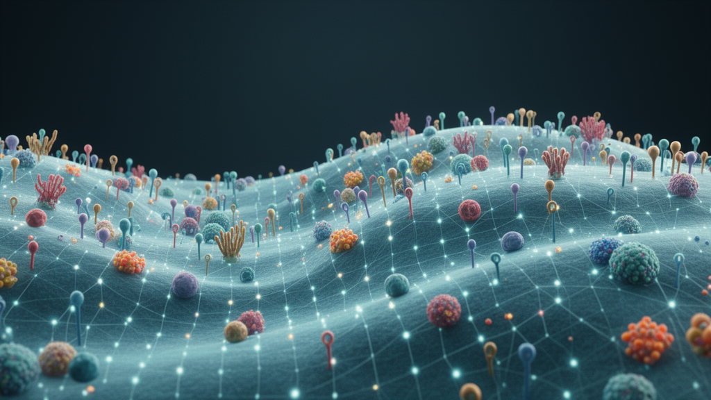
Researchers have mapped how proteins are arranged across the surface of human immune cells, revealing functional neighborhoods, including clusters not previously implicated in cell membrane functions.
The cell surface proteome, or surfaceome, governs processes essential for development, immunity and disease, yet its large-scale spatial organization had remained uncharted. To chart it, the team systematically mapped the surface proteome of human T lymphocytes and B lymphoblasts using proximity labeling of 85 antigens. The labeled proteins were then identified by data-independent acquisition mass spectrometry, producing a dense network of spatial relationships across the membrane.
The resulting map revealed functional modules on the cell surface. Some corresponded to known complexes, such as the T cell receptor, while others were clusters not previously implicated in cell membrane functions. The analysis also uncovered a surprise: mitochondrial proteins, normally thought of as residents of organelles inside the cell, were detected at the cell surface. The authors suggest these proteins may bear underappreciated roles at the plasma membrane, though the functional consequences of their surface localization remain to be worked out.
Beyond mapping, the team built a machine learning classifier trained on the proximity labeling data to predict associations between surface proteins. The classifier predicted surface protein associations, revealing the IL-10 receptor beta subunit's regulation of type I interferon signaling. The work also included three-dimensional network modeling that distinguished widely dispersed antigens from those localized to particular regions of the surface, indicating that active mechanisms organize the cell surface rather than proteins simply diffusing at random.
The authors present the study as a large-scale map of the human surfaceome and a resource for exploring its spatial and functional organization. Because the mapping focused on T lymphocytes and B lymphoblasts, the findings describe immune cell surfaces specifically, and the researchers note that the spatial patterns they observed hint at how cells may actively maintain or remodel the arrangement of their surface machinery. The dataset and predictive tools are intended to help other researchers identify surface protein associations and investigate the roles of proteins found in unexpected membrane locations.
Subject of Research: Spatial and functional organization of the human cell surface proteome in immune cells
Article Title: A nanoscale map of the human cell surface proteome reveals spatial and functional organization
Article References: Floyd, B. M., Schmidt, E. L., Till, N. A., Waldman, B. S., Yang, J. L., Liao, P., George, B. M., Carette, J. E., Flynn, R. A., & Bertozzi, C. R. (2026). A nanoscale map of the human cell surface proteome reveals spatial and functional organization. Nature Structural & Molecular Biology. https://doi.org/10.1038/s41594-026-01914-4
Image Credits: AI Generated
DOI: 10.1038/s41594-026-01914-4
Keywords: cell surface proteome, surfaceome, proximity labeling, mass spectrometry, T lymphocytes, B lymphoblasts, mitochondrial proteins, machine learning, interferon signaling, IL-10 receptor, plasma membrane, protein interactions
News Source: Drew Townsend. (October 28, 2026). Nanoscale map charts the spatial organization of the human cell surface. Scienmag.