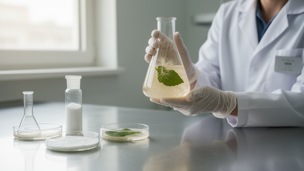
Zinc oxide nanoparticles grown with the help of a fungus harvested from hibiscus leaves can strip Staphylococcus aureus of two of its most damaging weapons, the enzymes lipase and hyaluronidase, while also choking the biofilms that make this bacterium so hard to treat. The study, published in Bioscience Nanotechnology, reports that nanoparticles biosynthesized by an endophytic fungus isolated from Hibiscus rosa-sinensis inhibited staphylococcal lipase with an estimated IC50 of roughly 380 micrograms per milliliter and hyaluronidase with an IC50 of about 330 micrograms per milliliter, and cut biofilm biomass by up to 68.7 percent at the highest concentration tested. In the same experiments the particles killed the bacterium outright, with a minimum inhibitory concentration of 1000 micrograms per milliliter and a time-dependent bactericidal effect that deepened over four and a half hours of exposure.
The work matters because of the direction it points rather than any immediate clinical claim. Methicillin-resistant Staphylococcus aureus, or MRSA, remains a global public health concern, driving skin and soft tissue infections, pneumonia and bloodstream infections, and its ability to form biofilms and deploy an arsenal of toxins and extracellular enzymes has complicated treatment strategies. The authors argue that targeting virulence mechanisms, rather than simply trying to kill the pathogen, may lower the selective pressure that fuels resistance development. Their nanoparticles, made through a biological synthesis route that avoids harsh chemical reagents, were designed as a probe of exactly that idea: an antivirulence material that also happens to be bactericidal at high enough doses.
The starting material was a fungus living quietly inside the tissues of Hibiscus rosa-sinensis, one of four Indian medicinal plants screened for endophytes in the study. Leaf segments from Withania somnifera, Hibiscus rosa-sinensis, Nerium oleander and Aegle marmelos were surface sterilized and cultured on potato dextrose agar, yielding four fungal isolates. Only one, designated HR1 and recovered from the hibiscus leaves, proved capable of efficiently reducing zinc sulfate into zinc oxide nanoparticles while also showing meaningful anti-staphylococcal activity, so it was carried forward for full characterization. Morphological examination, including colony texture, pigmentation and the distinctive branching conidiophores and chain arrangements seen under the microscope, placed the isolate in the genus Penicillium.
Species-level identity came from molecular work. The researchers amplified the internal transcribed spacer region of the fungal genome with universal primers ITS1 and ITS4, sequenced the product by Sanger methods, and compared it against the NCBI GenBank database, finding 99.50 percent similarity with Penicillium species. A maximum likelihood phylogenetic tree built in MEGA software clustered the strain, deposited under accession number PZ134185, closely with Penicillium citrinum, confirming the identification. The fungus was then grown in potato dextrose broth, the mycelia filtered out, and the cell-free culture filtrate mixed with zinc sulfate heptahydrate at pH 8. Within 12 to 24 hours at room temperature the mixture turned from light brown to dark grey and threw down a white precipitate, the visual signature of zinc ions being reduced to zinc oxide.
Characterization confirmed that the fungal metabolites had done their job. Ultraviolet-visible spectroscopy showed a distinct absorption peak between 260 and 280 nanometers, attributed to band-gap transitions in zinc oxide, a peak absent from both the precursor salt and the fungal filtrate alone. Fourier-transform infrared spectroscopy revealed a Zn-O stretching band between 430 and 500 wavenumbers along with signals for hydroxyl groups and protein capping, evidence that fungal biomolecules had coated and stabilized the particles. X-ray diffraction matched the hexagonal wurtzite structure of zinc oxide with no impurity peaks, and Scherrer analysis put the average crystallite size at 15 to 40 nanometers. Transmission electron microscopy showed predominantly spherical to quasi-spherical particles averaging 58.52 nanometers in Feret diameter, ranging from about 29 to 94 nanometers.
In suspension the particles behaved differently than on the dry grid. Dynamic light scattering gave a mean hydrodynamic diameter of 240.4 nanometers, larger than the TEM figure, which the authors attribute to a surface layer of fungal biomolecules. The polydispersity index was 0.2, indicating a narrow, essentially monodisperse size distribution, and the zeta potential of roughly minus 13 millivolts pointed to electrostatic repulsion between particles strong enough to keep the suspension stable without aggregation. The reaction mixture itself remained free of aggregation and discoloration for at least 48 hours, and the ultraviolet absorption signature persisted for several days, both signs that the biological capping was doing real work.
Against S. aureus the particles performed respectably in standard assays. In agar well diffusion tests, 1000 micrograms per milliliter of the nanoparticles produced a zone of inhibition of 20 plus or minus 1.13 millimeters, close to the 22 plus or minus 1.93 millimeters produced by the streptomycin positive control, while the fungal filtrate alone and sterile saline produced no zone at all. Tube dilution testing fixed the minimum inhibitory concentration at 1000 micrograms per milliliter, and colony-forming counts dropped to zero at that concentration, confirming a bactericidal rather than merely bacteriostatic effect. In time-kill experiments, absorbance fell sharply within the first hour, continued declining through 150 and 180 minutes, and reached its lowest point at 270 minutes, a profile the authors describe as time-dependent killing.
The antivirulence results were the study’s distinctive contribution. In crystal violet assays of biofilm biomass, inhibition was negligible at low nanoparticle concentrations, reached 31.9 percent at 800 micrograms per milliliter and peaked at 68.7 percent at 1000 micrograms per milliliter, a statistically significant, dose-dependent pattern. A parallel methylene blue reduction test, which tracks the metabolic activity of biofilm-embedded cells by measuring how quickly living bacteria decolorize the dye, showed a concentration-dependent delay in decolorization, corroborating that the particles were suppressing the viability and metabolic function of cells inside the biofilm, not just reducing biomass. On the enzyme front, qualitative plate assays showed that pre-incubating staphylococcal lipase or hyaluronidase with the nanoparticles abolished or sharply reduced the characteristic reaction halos, matching the effect of the standard inhibitors quercetin and tannic acid respectively.
Quantitative enzyme assays sharpened the picture. In a tributyrin tube assay, lipase inhibition climbed with nanoparticle concentration to a maximum of 65.60 percent at 1000 micrograms per milliliter, with the halfway point reached near 350 micrograms per milliliter, yielding the estimated IC50 of about 380 micrograms per milliliter. Hyaluronidase inhibition rose from 32.3 percent at 200 micrograms per milliliter to 67.5 percent at 1000 micrograms per milliliter, with an interpolated IC50 of 330 micrograms per milliliter, while tannic acid achieved 80 percent inhibition as an assay control. The authors suggest the mechanism may involve zinc ions interacting with catalytic serine and histidine residues in lipase, direct adsorption of nanoparticles onto the enzyme surface distorting the active site, or general conformational destabilization of the proteins, though they note these mechanistic explanations remain inferential.
Safety screening offered cautious reassurance within its narrow scope. When human red blood cells were incubated with the nanoparticles across the tested concentration range, hemolysis stayed below 2 percent even at the highest dose of 1000 micrograms per milliliter, well within standard hemocompatibility limits and far from the distilled-water positive control that produced complete lysis. The authors are careful about what this does and does not establish: the biocompatibility result supports the particles’ suitability for further biomedical investigation, but the study is entirely in vitro, and they explicitly state that detailed in vivo studies and mechanistic investigations are necessary before any clinical applicability can be claimed. What the work does demonstrate is a reproducible, biologically mediated route to stable zinc oxide nanoparticles from a common endophytic fungus, and evidence that those particles can simultaneously blunt the enzymatic virulence machinery, disrupt biofilm formation and kill S. aureus in laboratory assays, a combination the researchers present as a promising template for antivirulence strategies against drug-resistant staphylococcal infections.
Subject of Research: Biosynthesis of zinc oxide nanoparticles by an endophytic fungus and their inhibition of Staphylococcus aureus virulence enzymes and biofilm formation
Article Title: Green-synthesized ZnO nanoparticles from endophytic fungi inhibit virulence exoenzymes and biofilm formation in Staphylococcus aureus
Article References: Joshi, A. A., Songire, V. M., & Patil, R. H. (2026). Green-synthesized ZnO nanoparticles from endophytic fungi inhibit virulence exoenzymes and biofilm formation in Staphylococcus aureus. Bioscience Nanotechnology, 2(1), Article 22. https://doi.org/10.1186/s44331-026-00029-3
Image Credits: AI Generated
DOI: 10.1186/s44331-026-00029-3
Keywords: zinc oxide nanoparticles, green synthesis, endophytic fungi, Penicillium citrinum, Staphylococcus aureus, MRSA, biofilm, virulence enzymes, lipase, hyaluronidase, antivirulence, hemocompatibility
News Source: Roger Howard. (October 28, 2026). Fungal endophyte from hibiscus yields zinc oxide nanoparticles that curb Staphylococcus aureus virulence. Scienmag.