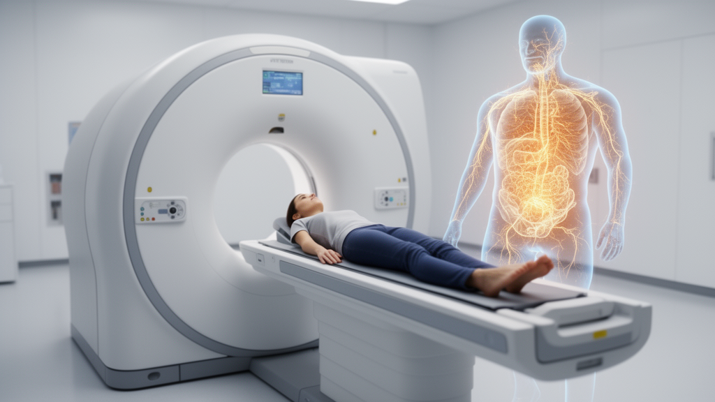
Photon-counting CT can detect and reliably quantify far smaller amounts of iodine than today’s dual-energy CT systems, particularly when radiation dose is low and the region of interest is small, according to a phantom study published in European Radiology Experimental. In head-equivalent and abdomen-equivalent imaging conditions, the photon-counting scanner achieved significantly lower limits of detection and quantification than a dual-source and a split-filter dual-energy system across most protocol combinations, with the largest advantages appearing in the low-dose settings that matter most for patients who need contrast media or radiation kept to a minimum.
The study, led by Rebecca Titternes and colleagues, is the first to apply the laboratory-science concepts of limit of detection, limit of quantification, and lower limit of the linear interval to a photon-counting CT system. These metrics, long established in clinical chemistry and molecular diagnostics and codified in Clinical and Laboratory Standards Institute guidelines EP17-A2 and EP06, describe in concentration terms exactly what an imaging system can and cannot do: the lowest iodine level distinguishable from background noise, the lowest level at which a region-of-interest measurement is quantitatively reliable, and the lowest level at which measurements remain proportional to the true iodine content. Until now, no study had compared these thresholds across photon-counting and dual-energy platforms.
The motivation is practical. Iodine maps from CT are widely used in neuro and abdominal imaging to quantify iodine uptake, characterize lesions, distinguish contrast extravasation from hemorrhage, and identify subtle enhancement. Clinicians frequently interpret these maps against concentration thresholds, so the central question is whether a given measurement can be trusted. Conventional image quality metrics such as contrast-to-noise ratio rest on assumptions about noise properties and reconstruction algorithms that limit their suitability for comparing different CT systems, especially when spatial resolution or noise texture differ. Model observer methods can overcome some of these shortcomings but typically focus on lesion detectability rather than quantification of a signal within a region.
To build a controlled comparison, the team constructed a 15 by 20 centimeter solid PMMA cylinder with radially drilled holes holding conical centrifuge tubes filled with iohexol diluted in distilled water to concentrations of 2, 1, 0.5, 0.25, 0.125, 0.0625, 0.03125, and 0 milligrams per milliliter. This phantom approximates the X-ray attenuation of a human head. To simulate abdominal imaging, the cylinder was placed inside an elliptical body annulus measuring 25 by 35 centimeters with a thickness of 15 centimeters. Three clinical Siemens Healthineers systems were tested: a dual-source SOMATOM Force, a split-filter SOMATOM X.ceed, and a photon-counting NAEOTOM Alpha.
Each system was evaluated under four protocol settings, a head and an abdominal examination protocol each imaged at a low and a high radiation dose, using manufacturers’ recommended protocols with adjusted CTDIvol values of 3 and 13 mGy for the abdomen and 15 and 50 mGy for the head. Every protocol was repeated 20 times per system. Iodine maps, derived from a three-material decomposition model with iodine, fat, and soft tissue as basis materials, were reconstructed with a Qr40 kernel, 1.5-millimeter slice thickness, and fields of view of 210 millimeters for brain and 360 millimeters for abdominal protocols. Iterative reconstruction was applied at varying levels, QIR levels 1 to 4 for the photon-counting system and ADMIRE levels 1 to 5 for the dual-energy systems.
A key methodological step was an independent calibration. Because vendor-provided calibrations vary across scanners, protocols, and reconstruction algorithms, the researchers mapped measured Hounsfield unit signals from regions of interest placed at the center of each test tube, averaged across selected slices and all 20 repeats, against the known iodine concentrations using linear least squares regression. This allowed all systems to be evaluated against the same true concentration, enabling system- and protocol-agnostic comparison. The limit of blank was estimated parametrically as the 95th percentile of the blank distribution, the limit of detection as the lowest concentration whose signal lower bound exceeded that threshold, and the limit of quantification by interpolating a precision profile to a target coefficient of variation of 20 percent, following CLSI EP17-A2.
The results favored photon-counting CT decisively in the clinically challenging conditions. For the low-dose abdominal protocol at 3 mGy with medium iterative reconstruction, the limit of detection for a 3-millimeter region of interest was 0.78 milligrams per milliliter for photon-counting CT, compared with 1.28 for split-filter CT and 1.42 for dual-source CT. For a 10-millimeter region, the corresponding values were 0.16, 0.26, and 0.57 milligrams per milliliter. Limits of quantification were also lower for the photon-counting system, and improvements were most pronounced in low-dose protocols, with detection-limit reductions of up to 68 percent in brain-equivalent and 45 percent in abdomen-equivalent conditions. The lower limit of the linear interval was similar across systems.
Statistical analysis using one-way Welch ANOVA with Games-Howell post hoc tests confirmed the pattern. At iterative reconstruction level 3, photon-counting CT showed significantly lower detection limits for 3-millimeter regions across all protocols, all p at or below 0.003, except the high-dose brain protocol where it matched the split-filter system. Quantification limits were significantly lower across all four protocols for the small regions. For 10-millimeter regions, the photon-counting advantage persisted in the low-dose abdominal and high-dose brain protocols, while performance converged with the dual-energy systems in the high-dose abdominal and low-dose brain settings. The photon-counting scanner also consistently showed the highest contrast response per milligram per milliliter and the lowest image noise.
The clinical context gives these numbers weight. Prior work cited in the study reports that dual-energy iodine quantification can differentiate benign from malignant renal lesions using thresholds around 0.5 milligrams per milliliter, that iodine density differences between ischemic and non-ischemic tissue in stroke patients are roughly 0.3 milligrams per milliliter, and that traumatic contusions show leakage ranging from 0.1 to 1.0 milligrams per milliliter. Against such values, the finding that both dual-energy systems exceeded the upper calibration range of 2 milligrams per milliliter for their quantification limit in small regions under low-dose abdominal protocols is striking, suggesting that small-lesion iodine measurements in low-dose dual-energy imaging should be interpreted with caution, while the photon-counting system maintained relatively lower quantification limits even under these conditions.
The authors are careful about scope. The phantom-based design is a simplified environment that does not capture tissue heterogeneity or anatomical complexity, the slice-wise analysis may retain residual correlation from overlapping helical reconstructions so that reported confidence intervals may be underestimated, and only a single vendor’s systems were tested. The results should be read as baseline estimates of detection and quantification performance under controlled conditions. Even so, the implications are concrete: under conditions resembling the phantom experiments, photon-counting CT could support detecting and quantifying similar lesions with less contrast media, or smaller regions at the same dose, and previous clinical studies have shown contrast volume reductions of roughly 17 to 30 percent in thoraco-abdominal and abdominal examinations with first-generation photon-counting CT without harming image quality. The authors suggest these findings may support future protocol optimization, such as iodine contrast or radiation dose reduction, pending clinical validation.
Subject of Research: Comparing iodine detection and quantification limits between photon-counting CT and dual-energy CT systems using a head and abdomen phantom
Article Title: Evaluation of iodine detection and quantification limits in photon-counting CT—a phantom study
Article References: Titternes, R., Poludniowski, G., Brismar, T. B., Nowik, P., & Thor, D. (2026). Evaluation of iodine detection and quantification limits in photon-counting CT—a phantom study. European Radiology Experimental, 10(1), Article 154. https://doi.org/10.1186/s41747-026-00824-w
Image Credits: AI Generated
DOI: 10.1186/s41747-026-00824-w
Keywords: photon-counting CT, dual-energy CT, iodine quantification, limit of detection, limit of quantification, phantom study, radiation dose, iterative reconstruction, iodine maps, contrast media, CT image quality, clinical chemistry metrics
News Source: Ophelia Keating. (October 29, 2026). Photon-counting CT outperforms dual-energy scanners at detecting tiny iodine traces. Scienmag.