Antibiotics save lives, but they can also strip the gut of the microbial communities that keep digestion and immunity running smoothly, leaving some patients with antibiotic-associated diarrhea. A research team in China now reports that a carefully paired probiotic combination—one wild-type strain of Lactiplantibacillus plantarum C8 and a genetically engineered version of the same strain overexpressing surface proteins with the LPxTG motif—outperformed either strain alone at restoring gut health in a mouse model of antibiotic-induced dysbiosis. The work, published in a Springer Nature journal covering agricultural products processing and storage, points to engineered microbial consortia as a possible route toward protecting the intestine during antimicrobial therapy.
The study’s central idea rests on a structural feature of many Gram-positive bacteria. LPxTG proteins are surface proteins covalently anchored to the bacterial cell wall by the enzyme sortase, and in L. plantarum they play a critical role in adhesion—the ability of a probiotic to attach to intestinal epithelial cells and establish stable colonization. Because colonization is a key determinant of probiotic efficacy, the researchers reasoned that boosting LPxTG display could strengthen the strain’s grip on the gut. They also drew on evidence that expression of these proteins is regulated by quorum sensing, a cell-density-dependent communication system that lets bacterial populations coordinate behavior, modulate adhesion capacity, and persist in response to environmental cues.
To build the engineered strain, the team identified signal peptides in the proteome of L. plantarum C8 using the NCBI and UniProt databases, then cloned the LPxTG motif protein gene together with a signal peptide gene into the pMG36e plasmid vector through homologous recombination. The construct was first amplified in chemically competent BL21 cells and then electroporated into competent L. plantarum C8 cells. The resulting recombinant strain, along with an empty-vector control, was cultured in MRS medium containing erythromycin for selection, activated over three generations, and finally co-cultured with the wild-type strain for 12 hours before the bacterial suspension was washed and standardized to an optical density at 600 nanometers of 1.00 ± 0.05 for animal dosing.
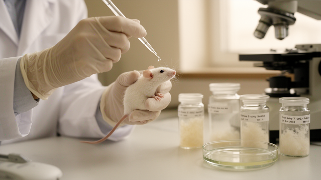
The animal model used 56 SPF-grade male KM mice, roughly six weeks old and weighing 25 ± 2 grams, obtained from a commercial supplier in Beijing. After a seven-day acclimatization, 48 mice received 300 mg/mL lincomycin hydrochloride by oral gavage twice daily for three consecutive days to induce intestinal dysbiosis, while eight mice served as an untreated blank control. The dysbiotic animals were then divided into six treatment subgroups of eight each: natural recovery with saline, wild-type L. plantarum C8 alone, empty-vector strain alone, recombinant strain alone, a co-culture of wild-type with empty-vector strain, and a co-culture of wild-type with the LPxTG-overexpressing recombinant strain. Over a 25-day experiment, the researchers tracked daily body weight, collected fecal samples, and on day 25 collected serum, colon, liver, and spleen tissues after euthanasia, with approval from the Ningbo University Animal Ethics Committee.
Body weight told the first part of the story. No mice died during the experiment, and all groups gained weight steadily during adaptation. Lincomycin treatment reversed that trend, producing a gradual weight decline while the blank control group continued to gain. During recovery, the natural-recovery group gained significantly less weight than any bacterial intervention group, and among the treated animals the wild-type strain improved recovery while the co-culture of wild-type and recombinant strains showed the most pronounced effect—an outcome the authors attribute to rapid intestinal colonization mediated by LPxTG-dependent adhesion of the engineered strain.
Colonic anatomy and organ indices followed a similar pattern. Colon length in the natural-recovery group fell to 5.43 ± 0.86 centimeters, a hallmark of colonic inflammation, whereas all probiotic-treated groups recovered lengths between 7.06 and 8.37 centimeters, approximating or exceeding the blank control value of 7.26 ± 0.63 centimeters. The liver index, which was significantly elevated above 5 percent in the saline-treated recovery group—signaling increased hepatic metabolic burden—returned to levels comparable to the blank control in the wild-type, recombinant, and co-culture groups, while empty-vector strains and their co-culture showed weaker recovery, possibly because plasmid maintenance imposes a metabolic burden on the bacteria. Spleen indices, elevated in the natural-recovery group as a sign of immune dysregulation, recovered best in the recombinant and co-culture groups.
Histology and molecular assays added mechanistic depth. Hematoxylin and eosin staining revealed intact colonic architecture in controls but disrupted intestinal villi, abnormal goblet cell morphology, and substantial inflammatory infiltration in the natural-recovery group; all interventions improved these lesions, with the co-culture group’s tissue most closely resembling normal structure. Serum cytokine measurements showed that antibiotic treatment raised the pro-inflammatory cytokines IL-6 and TNF-α by 98.34 percent and 83.99 percent respectively, while cutting the anti-inflammatory cytokine IL-10 by 73.43 percent, all statistically significant. Probiotic treatment lowered IL-6 and TNF-α to 132.00 and 131.83 pg/mg in the wild-type-plus-recombinant co-culture group—the lowest values among all interventions—while raising IL-10 to 226.47 pg/mg, the highest level recorded.
Tight junction proteins told a parallel story. Immunohistochemistry showed that antibiotic exposure reduced colonic expression of ZO-1 and Occludin, proteins essential for sealing the paracellular space between epithelial cells, to only 14.77 percent and 15.42 percent of blank-control levels in the natural-recovery group. All probiotic interventions significantly upregulated these proteins, and the co-culture group achieved the strongest restoration, reaching 31.54 percent and 31.33 percent of control levels for ZO-1 and Occludin—roughly a 2.1-fold improvement over natural recovery and significantly better than the 22.99 to 28.11 percent range seen with single-strain treatments. The authors link this barrier repair to reduced translocation of pro-inflammatory microbial products into circulation, which they connect to the observed normalization of liver and spleen indices.
Gut microbiota sequencing underscored the ecological dimension. The blank control group showed the highest Chao1 and Shannon alpha-diversity indices, while the natural-recovery group suffered significant reductions, confirming severe disruption by lincomycin. All probiotic groups ameliorated the dysbiosis, but the wild-type-plus-recombinant co-culture recovered diversity to the point that its index showed no statistically significant difference from the blank control. Principal coordinates analysis confirmed that the antibiotic significantly reshaped beta-diversity (P = 0.001, R = 0.193), and the co-culture group’s community structure most closely resembled the healthy control. At the phylum level, Bacteroidota abundance, which dropped 30.51 percent in the natural-recovery group, rebounded by 50.96 percent in the co-culture group, while a more modest 11.37 percent rise in Firmicutes helped restore the Firmicutes-to-Bacteroidota ratio toward control values. At the genus level, Bacteroides rose 49.55 percent and Lactobacillus surged 331.34 percent relative to natural recovery.
The authors propose that the synergy arises from three coordinated actions of the LPxTG proteins and signal peptides: enhanced adhesion prolongs colonization of both strains; overexpressed signal peptides persistently activate histidine kinase, amplifying quorum-sensing signals and stimulating antimicrobial production by the wild-type strain; and LPxTG-mediated bacterial aggregation through homologous or heterologous interactions supports biofilm microcolony formation, giving the consortium a protective niche against stress. In this picture the engineered strain acts as a structural scaffold to which wild-type bacteria adhere, and sortase A—the LPxTG-anchoring enzyme—has previously been shown in L. plantarum C8 to regulate adhesion, biofilm formation, and AI-2-dependent quorum sensing, the most prevalent interspecies signal in the gut. The team also notes a caution: monoculture of an overexpressing engineered strain may impose excessive competitive pressure, so pairing it with the wild-type parent creates an ecological buffer that preserves niches for indigenous microbiota recolonization. As a mouse study with a single strain pair and a defined antibiotic challenge, the work establishes a mechanistic foundation rather than a clinical result, but it offers a concrete design principle—engineered surface-protein display combined with wild-type partnership—for future probiotic strategies against antibiotic-induced intestinal dysbiosis.
Subject of Research: Engineered LPxTG surface protein overexpression in Lactiplantibacillus plantarum C8 co-cultures for alleviating antibiotic-associated diarrhea in mice
Article Title: LPxTG proteins mediate microbial interactions to modulate Lactiplantibacillus plantarum C8 in alleviating antibiotic-associated diarrhea
Article References: Liang, J., Liu, J., Zhang, A., Zheng, S., Jiang, T., Yang, L., Qi, X., Zeng, X., Guo, Y., Pan, D., Zhang, T., & Wu, Z. (2026). LPxTG proteins mediate microbial interactions to modulate Lactiplantibacillus plantarum C8 in alleviating antibiotic-associated diarrhea. Agricultural Products Processing and Storage, 2(1), Article 42. https://doi.org/10.1007/s44462-026-00076-2
Image Credits: AI Generated
DOI: 10.1007/s44462-026-00076-2
Keywords: antibiotic-associated diarrhea, Lactiplantibacillus plantarum C8, LPxTG proteins, gut microbiota, probiotics, quorum sensing, tight junction proteins, intestinal barrier, mouse model, dysbiosis, IL-10, sortase
News Source: Morgan Morrow. (October 28, 2026). Engineered Probiotic Co-Culture Eases Antibiotic-Linked Diarrhea in Mice. Scienmag.