Segmenting individual thigh muscles on MRI has long been a stubborn problem for automated image analysis. The muscles share borders with little contrast between them, so while whole-thigh compartment segmentation can reach Dice scores of roughly 0.96, accuracy for individual muscle groups has typically dropped to around 0.83 to 0.91. A new study published in European Radiology Experimental reports that a pre-trained foundation model, fine-tuned on a comparatively small set of manually annotated scans, can segment the major thigh muscle groups with Dice scores ranging from 0.89 to 0.97 — performance the authors describe as comparable to purpose-built networks, without training a dedicated architecture from scratch.
The research team, led by Katharina Ziegeler and Gabrielle Hoyer with senior author Thomas M. Link, worked with data from the Osteoarthritis Initiative (OAI), a publicly available, multi-center observational study of people with or at risk of knee osteoarthritis. A subset of OAI participants underwent 3.0-Tesla MRI of both thighs. The researchers used manual segmentations of thigh muscle groups on axial T1-weighted spin echo images from 364 participants: 106 for training and validation, split 80/20, and 258 held-out testing scans that were never used for training or fine-tuning.
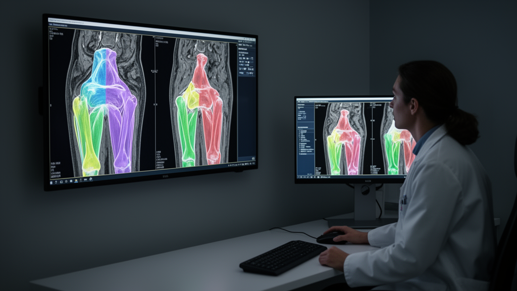
The pipeline itself has two stages. First, a You Only Look Once (YOLO) object detection model, trained specifically for this task, generates bounding boxes and class labels for each major muscle group on the axial images. Those boxes then serve as spatial prompts for the Segment Anything Model (SAM) from Meta, fine-tuned on the thigh muscle data using a Vision Transformer ViT-B backbone. SAM combines an image encoder, a prompt encoder that processes the bounding box inputs, and a mask decoder that produces the binary segmentation masks. The authors emphasize that SAM does not work reliably on muscle MRI out of the box; its advantage lies in reaching task-specific accuracy through fine-tuning on modest domain-specific data rather than zero-shot performance.
The images covered the mid-thighs, spanning approximately 7.5 centimeters starting 10 centimeters proximal to the right femoral epiphysis, with fifteen slices acquired and the central seven used as the unit of observation to limit edge-slice signal inhomogeneity. Five muscle groups were analyzed: quadriceps, hamstrings, adductors, gracilis, and sartorius. Manual reference masks were created in ITK-SNAP by three board-certified radiologists, who corrected initial proposed masks slice by slice. On the testing set, Dice scores were 0.97 for quadriceps, 0.96 for hamstrings, 0.89 for adductors, 0.93 for gracilis, and 0.94 for sartorius. Relative to ground-truth muscle volumes, mean bias ranged from -0.1 percent for the quadriceps to 4.7 percent for the gracilis.
A central question of the study was whether demographic factors bias segmentation accuracy — a concern given documented racial disparities in cardiac MRI segmentation and demographic performance gaps in musculoskeletal radiograph segmentation. In separate linear regression models, the only significant association was female sex, which was linked to marginally lower Dice scores (beta -0.01, 95% CI: -0.02, -0.01, p < 0.001). This association remained significant after adjustment for age and BMI (beta -0.01, p = 0.002). The authors suggest a possible biological explanation: women have thicker subcutaneous fat and smaller muscle volumes, which may make the segmentation task harder. Reassuringly, no significant associations were found for race, BMI, or age — the latter somewhat contrary to the team’s expectation, since thigh muscle fat content increases with age and reduces tissue contrast.
The second focus was on what visually flawed segmentations actually do to the numbers. Two radiologists graded every segmentation in the testing set as having no issue, a minor issue, or a major issue, adapted from an existing quality assurance score. Intra-reader agreement was almost perfect, with weighted Cohen’s kappa between 0.93 and 0.99, and inter-reader agreement was substantial to almost perfect, ranging from 0.79 to 0.88. Failure of segmentation was rarest in the quadriceps (2.3 percent of cases) and most frequent in the adductors (14.3 percent).
The comparison between flagged and unflagged cases yielded a striking dissociation. Mean Dice scores were significantly lower in cases with major issues for the hamstrings, adductors, gracilis, and sartorius — for example, 0.90 versus 0.84 for the adductors — but the differences ranged only from 0.016 to 0.068, which the authors judged too small to be clinically meaningful. More importantly, excluding the error cases did not meaningfully improve the volumetric measurements. Averaged across all muscle groups, exclusion improved mean bias by just 0.06 mL. For the quadriceps, mean bias shifted from -0.34 to -0.17 mL; for the hamstrings, from 7.87 to 7.31 mL; while for the adductors the near-zero bias actually shifted marginally from +0.09 to -0.53 mL. All changes were tiny relative to the Bland–Altman limits of agreement, which reached approximately ±29 mL for the adductors.
The reason for this dissociation, the authors explain, is that visual assessment and Dice capture the magnitude of overlap error, whereas mean bias reflects signed deviations that can partially offset across cases — so the visually flagged cases were not necessarily the volumetrically most deviant ones. This has a practical downside: a simple data-driven outlier rule based on Dice scores (flagging cases with a z-score below -2 within each muscle group) recovered only a small fraction of the radiologist-flagged errors, with recall of 0.17, 0.12, and 0.14 for quadriceps, hamstrings, and adductors respectively, and precision of at most 0.50 in all groups. Qualitative severity grading itself discriminated the most severe failures reasonably well (AUC 0.62 to 0.94 for none-versus-major comparisons) but performed at or near chance for distinguishing minor issues from unflagged cases (AUC 0.47 to 0.61), and was only weakly associated with overlap overall (pooled Spearman ρ = -0.16).
The team is careful about scope and limitations. The analysis was a post-hoc study on a single cohort, and the pipeline was not tested on external datasets where performance might be lower. The quality assessment did not separately evaluate left and right thighs, even though errors were often unilateral. Only muscle volume was evaluated as a downstream metric — not intramuscular fat, muscle shape, or texture parameters. The ViT-B variant of the original SAM was used rather than a larger backbone or the later-released SAM 2, and no systematic data augmentation was explored. Comparisons against other methods were indirect, differing in cohort, sequence, and reference standard, and a head-to-head benchmark against a trained nnU-Net remains future work. The authors also caution that while the findings are reassuring for research contexts focused on group statistics, errors may matter more when segmentations inform decisions about individual patients, such as predicting operative success before knee replacement surgery.
The broader context gives the work its urgency. Knee osteoarthritis is the leading cause of disability worldwide, with an estimated national cost of $45 billion annually in the United States, and medications proven to slow the disease are still missing. Muscle weakness predicts both incidence and progression of radiographic knee osteoarthritis, making objective assessment of muscle quality essential for understanding disease mechanisms and evaluating treatments aimed at building muscle. By showing that a foundation model can be fine-tuned on a modest task-specific dataset to achieve accuracy approaching whole-compartment segmentation, and that radiologist-identified visual errors introduce only negligible degradation of volumetric metrics, the study suggests that rigorous per-case quality control after initial validation may not be necessary for scaled research datasets — while simultaneously revealing that such errors are difficult to catch with statistical detection methods alone.
Subject of Research: Validation of a foundation-model pipeline for automated thigh muscle segmentation on MRI and the impact of segmentation quality on volumetric metrics
Article Title: Automated thigh muscle segmentation using foundation models: validation and impact of segmentation quality
Article References: Ziegeler, K., Hoyer, G., Liu, F., Kreutzinger, V., Akkaya, Z., Joseph, G. B., Lynch, J. A., Pedoia, V., Majumdar, S., & Link, T. M. (2026). Automated thigh muscle segmentation using foundation models: validation and impact of segmentation quality. European Radiology Experimental, 10(1), Article 157. https://doi.org/10.1186/s41747-026-00822-y
Image Credits: AI Generated
DOI: 10.1186/s41747-026-00822-y
Keywords: thigh muscle segmentation, foundation models, Segment Anything Model, MRI, Osteoarthritis Initiative, Dice score, deep learning, muscle volume, quality assurance, demographic bias, YOLO, radiology
News Source: Blake Davidson. (October 29, 2026). Foundation model segments thigh muscles on MRI with near-bespoke accuracy. Scienmag.