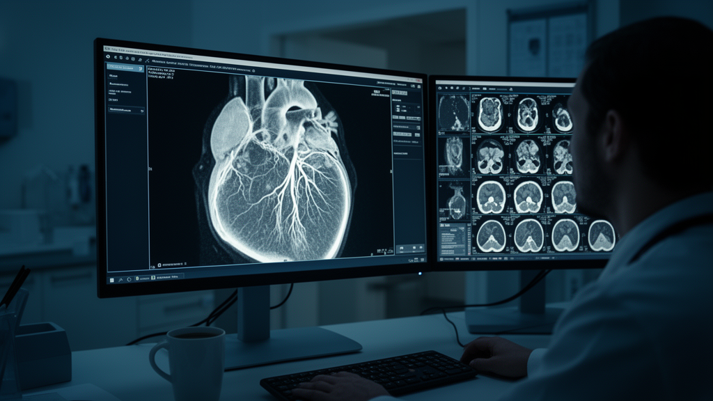
A real-world reduction in the amount of iodinated contrast injected during coronary CT angiography cut the administered volume and total iodine load by nearly a quarter, but it also left a small minority of patients with coronary artery attenuation below the accepted diagnostic threshold, according to a retrospective before-and-after study from a radiology centre in Cairo, Egypt. The findings, published in the Egyptian Journal of Radiology and Nuclear Medicine, offer a candid look at what happens when a contrast-sparing protocol moves out of the research setting and into routine practice.
Coronary CT angiography, or CCTA, depends on intravenous iodinated contrast to opacify the coronary lumen brightly enough for the scanner to delineate the vessel interior. Reducing the contrast volume per examination is an attractive exposure-reduction goal: it lowers the iodine load each patient receives, and it matters practically in settings where contrast supply and cost are genuine constraints. Whether attenuation can be preserved above the diagnostic threshold of 300 Hounsfield units, a benchmark set by the Society of Cardiovascular Computed Tomography, under everyday non-randomized implementation is far less well documented than in purpose-built low-dose trials.
The study team identified 200 CCTA examinations from radiology reports as potentially eligible and, after excluding 106 — 44 for body weight above 120 kilograms, 12 for beta-blocker administration, 27 for coronary calcification or calcium scoring without significant stenosis, and 23 for motion artefact — analysed 94 CT-angiographically normal examinations. Fifty-one were performed under the standard pre-reduction protocol and 43 under the reduced-volume protocol introduced afterwards. All scans were acquired on a third-generation dual-source Siemens SOMATOM Force scanner with automated tube-voltage selection via CARE kV, tube-current modulation via CARE Dose4D, retrospective ECG gating and a fixed 32-centimetre scan length, so the acquisition hardware and gating mode were held constant across both eras.
Omnipaque at 350 milligrams of iodine per millilitre was power-injected with bolus tracking on the ascending aorta, a 90 HU trigger threshold and a 5-second post-trigger delay, with a 40-millilitre saline chaser in both groups. The average contrast volume fell from 63.4 millilitres to 49.0 millilitres, an absolute drop of 14.5 millilitres and a relative reduction of 22.8 percent, mirrored by an identical 22.8 percent fall in total iodine mass, from 22.20 to 17.13 grams of iodine. Crucially, the iodine delivery rate was essentially unchanged at 1.69 grams of iodine per second in both groups, meaning the intervention reduced the total iodine load rather than the pace at which iodine was delivered. Extrapolated to 100 examinations, the observed mean reduction corresponds to roughly 1,448 millilitres of contrast and 507 grams of iodine saved.
The attenuation cost of that saving was measurable. Mean attenuation in the left main trunk fell from 520.4 to 451.9 HU, a 13.2 percent decrease, and mean attenuation in the proximal right coronary artery fell from 500.2 to 418.1 HU, a 16.4 percent decrease, both statistically significant. Because the diagnostic threshold is a per-patient criterion rather than a group-average one, the researchers also counted individual examinations: 41 of 43 post-reduction scans, or 95.3 percent, reached at least 300 HU in the left main trunk, and 39 of 43, or 90.7 percent, did so in the right coronary artery. In other words, two post-reduction examinations fell below the threshold at the left main trunk and four at the right coronary artery, a detail that reliance on group means alone would have obscured. In the pre-reduction group, every one of 51 examinations met the threshold in the left main trunk and 50 of 51 did so in the right coronary artery.
To account for measurable confounders, the team fitted a multivariable linear model examining attenuation as a function of protocol epoch, tube voltage, body weight, heart rate and iodine delivery rate. Protocol epoch remained independently associated with lower attenuation after adjustment: a 75.7 HU decrease per post-reduction epoch at the left main trunk and an 87.8 HU decrease at the right coronary artery, both with confidence intervals excluding zero. Heart rate was independently and inversely associated with left main trunk attenuation, while tube voltage showed only a non-significant inverse trend and neither weight nor iodine delivery rate was independently associated. The authors are careful to note that this model demonstrates persistence of an association, not causality, and cannot exclude confounding by unmeasured variables such as reconstruction parameters.
Radiation dose was recorded as a secondary, co-temporal observation. Mean dose-length product was 1,158.2 milligray-centimetres in the pre-reduction group versus 1,072.1 in the post-reduction group, a 7.4 percent difference that did not reach statistical significance, and mean CTDIvol was similarly unchanged. The authors stress that contrast volume has no direct physical relationship to dose-length product, so any dose difference would require explanation by acquisition-parameter changes rather than by the contrast protocol itself; since all 94 examinations used retrospective gating in both eras, a shift in gating mode cannot explain the difference, but the dynamically selected tube voltage was not statistically equivalent by design, so a contribution from the kVp distribution cannot be excluded. The study was not designed to isolate contrast-protocol effects on radiation dose.
The limitations are substantial and the authors enumerate them candidly. The retrospective, non-concurrent, before-and-after design with sequential, non-randomized groups invites confounding by temporal changes in referral patterns, operator practice or unmeasured acquisition factors, and no sample-size or power calculation was performed. Attenuation was measured by a single reader with no inter- or intra-observer reproducibility assessment and no documented blinding to protocol epoch. No image noise, signal-to-noise ratio, contrast-to-noise ratio or blinded subjective image-quality scores were collected, so mean or per-patient Hounsfield units remain a necessary but insufficient surrogate for overall diagnostic image quality. The cohort was restricted to CT-angiographically normal coronary arteries, with high-weight, motion-degraded and calcium-positive examinations excluded, so the findings cannot be generalized to patients with coronary artery disease or elevated body weight, and no diagnostic-accuracy analysis against invasive angiography was performed.
Equally important is what the study does not claim about the kidneys. Although reducing iodine load is plausible on mechanistic grounds as a way of lowering theoretical contrast-associated renal risk, this study measured no renal endpoint whatsoever — no creatinine, no estimated glomerular filtration rate and no post-contrast or contrast-associated acute kidney injury outcome — and therefore cannot demonstrate any protective effect. The authors place their work in the context of contemporary controlled evidence finding little or no excess acute kidney injury risk attributable to intravenous contrast at CT doses once confounding by illness severity is accounted for, in contrast to older registries of intra-arterial contrast during percutaneous coronary intervention, whose risk models are not directly transferable to intravenous CCTA administration. Nor was the study designed with a pre-specified non-inferiority margin, so the results must not be read as formal proof of equivalence between protocols.
What remains is a carefully bounded but practically useful result: in routine, non-academic practice on a modern dual-source scanner, a 22.8 percent cut in contrast volume and iodine mass kept mean coronary attenuation above the 300 HU threshold in both protocol eras and kept more than 90 percent of individual post-reduction examinations above it at both measured sites, while the iodine delivery rate stayed constant. The authors conclude that these findings support the feasibility of meaningful contrast and iodine-load reduction in everyday CCTA, while indicating that per-patient attenuation monitoring — particularly in the right coronary artery, where the shortfall was concentrated — and prospective evaluation of renal and image-quality endpoints are warranted before any broader claims of clinical benefit can be made.
Subject of Research: Effects of a reduced iodinated contrast volume protocol on coronary artery attenuation in coronary CT angiography
Article Title: Reduced contrast volume in coronary CT angiography: effects on coronary attenuation
Article References: El Naqeeb, P. A. A., Shafaa, M. W., Ibrahim, A. M., Hamdy, A., & Ismail, A. S. (2026). Reduced contrast volume in coronary CT angiography: effects on coronary attenuation. Egyptian Journal of Radiology and Nuclear Medicine, 57(1), Article 215. https://doi.org/10.1186/s43055-026-01882-1
Image Credits: AI Generated
DOI: 10.1186/s43055-026-01882-1
Keywords: coronary CT angiography, iodinated contrast, contrast volume reduction, coronary attenuation, radiation dose, Hounsfield units, dual-source CT, retrospective study, iodine load, image quality, SCCT threshold, protocol optimization
News Source: Ophelia Keating. (October 29, 2026). Lower contrast doses in coronary CT keep most scans diagnostic, study finds. Scienmag.