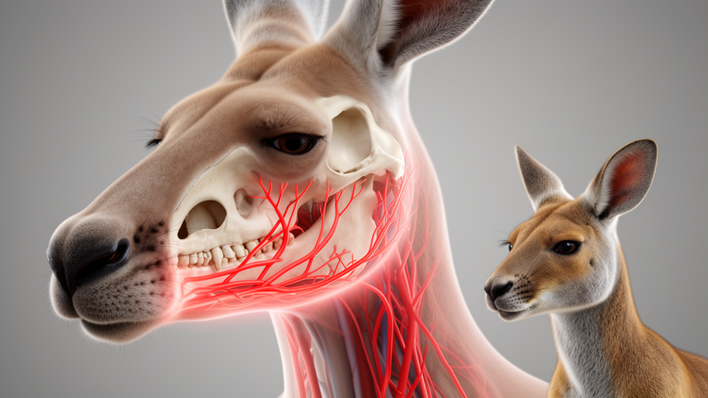
The red kangaroo and the red-necked wallaby, two Australian marsupial species commonly kept in zoological gardens, share a largely similar pattern of arterial supply to the face, according to a new anatomical study published in BMC Veterinary Research. The work, led by Maciej Zdun and Aleksander F. Butkiewicz, set out to chart in detail how blood reaches the facial region in these macropodids, an area the authors say has remained poorly documented even as exotic animal veterinary medicine expands.
The motivation is practical as much as academic. As the authors note in the paper’s background, cerebral vascularisation in these species has already been described, but data on the arteries supplying the face, which matter directly for surgery and dentistry, remain limited. Veterinarians treating kangaroos and wallabies in zoological settings therefore lack a reliable anatomical reference for procedures involving the lips, jaw, nasal cavity and oral structures.
To close that gap, the researchers assembled a substantial sample: 18 adult red kangaroos and 35 adult red-necked wallabies, all obtained from zoological gardens. Rather than relying on a single preparation technique, they applied three complementary methods. Latex was injected into the common carotid arteries to fill and reveal the arterial tree, corrosion casting with acrylic resin produced durable three-dimensional replicas of the vessels, and angio-CT imaging was performed after administration of barium sulphate as a contrast agent.
The resulting maps confirm the basic plan of the head’s blood supply in both species. The head is supplied by paired common carotid arteries, which divide at the level of the first cervical vertebra, C1, into the internal and external carotid arteries. From there, the main branches of the external carotid artery include the lingual, facial, caudal auricular and superficial temporal arteries.
The maxillary artery emerges as a key distributor. Before entering the maxilla, where it supplies the teeth and nasal structures, it gives off branches serving the masticatory muscles, the dura mater, the oral cavity, the nasal cavity and the palate. This branching sequence traces how a single major vessel underpins blood delivery to chewing, sensory and dental structures across the midface.
Several findings stand out for their comparative value. In both species, the lingual artery arises directly from the external carotid artery, and the submental artery forms a common trunk with the glandular branch. The lips, meanwhile, receive blood from branches of the facial, infraorbital and mental arteries, a distribution the authors highlight as differing from the patterns reported in ruminants and perissodactyls, the hoofed groups whose facial anatomy has historically anchored veterinary teaching.
One absence proved as informative as the structures found: the stapedial artery was not observed in either species.
Overall, the authors conclude that the arterial pattern was largely similar in the two species, suggesting a shared vascular blueprint within these macropodids despite their differences in size. The team positions the findings as a foundation for further anatomical, physiological and veterinary research on macropodids, and as potentially relevant to clinical procedures involving the head. Because the study examined animals from zoological collections and used post-mortem vascular preparations rather than living patients, its direct clinical application will depend on how these anatomical maps are tested in practice, but for veterinarians operating on the faces of kangaroos and wallabies, a detailed reference now exists where previously only limited data were available.
Subject of Research: The arterial vascularisation of the facial region in red kangaroos and red-necked wallabies
Article Title: Branches of the external carotid artery in the red kangaroo and the red-necked Wallaby
Article References: Zdun, M., & Butkiewicz, A. F. (2026). Branches of the external carotid artery in the red kangaroo and the red-necked Wallaby. BMC Veterinary Research. https://doi.org/10.1186/s12917-026-06049-8
Image Credits: AI Generated
DOI: 10.1186/s12917-026-06049-8
Keywords: red kangaroo, red-necked wallaby, external carotid artery, macropodids, veterinary anatomy, maxillary artery, corrosion casting, angio-CT, marsupials, zoological medicine, Branches, external
News Source: Drew Townsend. (October 28, 2026). Kangaroo and wallaby facial artery map revealed in new anatomical study. Scienmag.