Human umbilical cord mesenchymal stem cells, delivered through a simple tail-vein infusion, substantially repaired carbon tetrachloride-induced chronic liver injury in rats, according to a study published on 18 March 2026 in the journal Cell Regeneration. After three months of twice-weekly carbon tetrachloride injections had driven up liver enzymes, distorted hepatic architecture, and pushed the animals toward steatosis and fibrosis, three weeks of stem cell treatment measurably turned the picture around. Serum alanine aminotransferase, aspartate aminotransferase, and total bilirubin fell significantly, while total protein and albumin rebounded toward healthy levels. Under the microscope, hepatocyte steatosis receded and fibrous tissue hyperplasia all but vanished around vessels. The researchers then turned to targeted metabolomics to ask what, chemically, the cells had changed, and the answer pointed toward a specific corner of metabolism: propanoate metabolism, along with shifts in bile acid and caffeine-related metabolites.
The experimental design was deliberately layered. In cell culture, the team used THLE-2 human hepatic epithelial cells divided into blank control, carbon tetrachloride-treated, and carbon tetrachloride plus exosome groups, where the exosomes were vesicles secreted by the human umbilical cord mesenchymal stem cells. In parallel, 24 healthy specific-pathogen-free Sprague-Dawley male rats, ten weeks old and weighing roughly 400 grams, were randomly assigned to healthy, carbon tetrachloride, and carbon tetrachloride plus stem cell groups, eight animals per group. The model animals received intraperitoneal injections of 50 percent carbon tetrachloride in olive oil at 3 milliliters per kilogram twice weekly for three months, a standard way to provoke persistent hepatic inflammation and fibrosis. The treatment group then received tail-vein infusions of 1 × 10⁶ stem cells per milliliter twice weekly for three weeks, while the model controls received phosphate-buffered saline instead. Serum, liver tissue, and body mass were tracked throughout, and histopathology relied on hematoxylin-eosin and Masson trichrome staining to visualize cellular injury and collagen deposition, respectively.
Survival data underscored how punishing the toxin regimen was. All eight healthy rats survived the full protocol with glossy coats, normal appetite, and steady weight gain. In the untreated carbon tetrachloride group, only five of eight rats lived to the endpoint, and the survivors were lethargic with rough fur and diminished appetite. In the stem cell group, seven of eight survived. Body mass declined in both toxin-exposed groups, but the loss was significantly smaller in the treated animals, and the liver-to-body-weight ratio differed significantly between the two toxin groups even though absolute liver mass did not.
Biochemistry reinforced that impression. Compared with healthy controls, the carbon tetrachloride group showed significantly elevated alanine aminotransferase, aspartate aminotransferase, and total bilirubin, alongside significantly depressed total protein and albumin, confirming that the model had produced genuine liver dysfunction. After three weeks of stem cell therapy, the three injury markers dropped significantly and the two synthetic-function markers recovered to near-healthy values. Histology told the same story from the tissue side. Hematoxylin-eosin sections from the model group revealed hepatocyte necrosis, extensive vacuolar steatosis, nuclear fragmentation, disrupted hepatic cords, sinusoidal congestion, activated Kupffer cells, and dense inflammatory infiltration, while Masson staining exposed perisinusoidal fibrosis. In the treated group, hepatocyte numbers rose markedly, steatosis receded, congestion was minimal, and fibrous proliferation was confined to minor perivascular deposits.
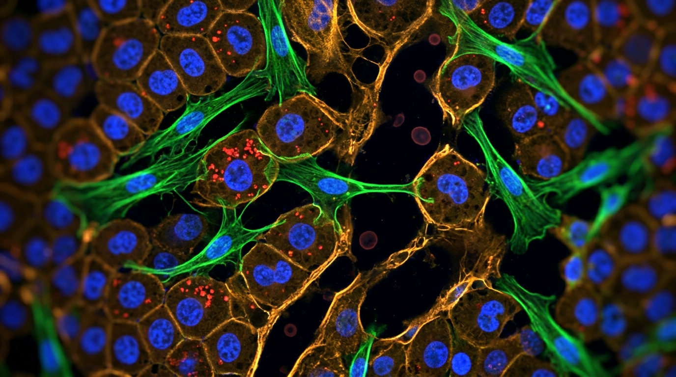
The cell-culture arm isolated one plausible mechanism. Exosomes, membrane-bound vesicles that inherit anti-inflammatory and tissue-regenerative properties from their parent stem cells, were applied to THLE-2 hepatocytes damaged by carbon tetrachloride. Cell viability, measured by CCK-8 assay, was significantly restored in the exosome-treated group, with the protective effect most evident at a carbon tetrachloride concentration of 16 millimoles per liter; at 18 millimoles per liter, no group fared well and the protective effect disappeared. Levels of aspartate aminotransferase, alanine aminotransferase, and malondialdehyde, a lipid peroxidation product, were significantly elevated by the toxin and significantly reduced by exosome co-treatment. Transmission electron microscopy showed that toxin exposure shrank mitochondria, vacuolized them, and fragmented their cristae, while exosome treatment preserved mitochondrial and cristae structure, though not fully to blank-control levels. Confocal microscopy with PKH26-labeled exosomes and MitoTracker Green-labeled mitochondria revealed clear spatial overlap, and three-dimensional Z-stack imaging confirmed that the vesicles physically localized to mitochondrial sites inside the cells.
Metabolomics supplied the chemical fingerprint. In the cellular targeted metabolomics assay, 48 metabolites differed significantly between the carbon tetrachloride and carbon tetrachloride plus exosome groups, with 33 increased and 15 decreased after exosome treatment. In the rat serum analysis, which profiled 16 samples across healthy, model, and treatment groups, 41 significant differential metabolites emerged between the model and treatment groups. Relative to the treated animals, the untreated model group showed significant upregulation of glycochenodeoxycholic acid, hydroxymethylbenzene, guanosine, and deoxyguanosine, and significant downregulation of sebacic acid, succinic acid, 4-methylpentanedioic acid, adipic acid, oxidized glutathione, and 2-hydroxybutyric acid. KEGG pathway enrichment pointed to propanoate metabolism, caffeine metabolism, and aromatic compound degradation as the dominant altered pathways. Notably, glycochenodeoxycholic acid and sebacic acid appeared in both the cellular and animal datasets with matching directional changes, lending cross-model consistency to the metabolomic signal.
Two metabolites stood out for sheer magnitude. In the treatment group, 3′-sialyllactose, a milk-derived oligosaccharide with documented anti-inflammatory and anti-apoptotic properties in prior literature, rose 9.13-fold relative to the model group, and aminophylline rose 7.71-fold. The authors connect 3′-sialyllactose to suppression of interleukin-1, interleukin-6, nitrite, prostaglandin E2, and cyclooxygenase-1 in stimulated cells, and to inhibition of hepatocyte apoptosis through downregulation of the pro-apoptotic protein Bax and upregulation of the anti-apoptotic protein B-cell lymphoma 2. Separately, oxidized glutathione was significantly upregulated and xanthine significantly downregulated in the treated group, a pattern the authors interpret as a shift toward better redox buffering, since glutathione peroxidase 4 consumes reduced glutathione to neutralize lipid hydroperoxides and produces oxidized glutathione in the process. These are associative observations from a targeted metabolomic platform, not proof of direct causation, but they align with the histological improvement.
The propanoate metabolism finding carries the most mechanistic weight. Propionic acid and propionate serve as major gluconeogenic substrates in hepatic metabolism and have been linked in prior research to enhanced insulin sensitivity and improved glucose and lipid handling. In this study, three differential metabolites enriched in the propanoate pathway, 2-hydroxybutanoic acid, succinic acid, and methylmalonic acid, were all upregulated in the stem cell group relative to the model group. The authors propose that this upregulation helps explain the observed reduction in hepatocyte steatosis, noting that propionate has been shown in earlier work to inhibit hepatic lipid synthesis and promote lipid transport and oxidation through activation of the AMPK-PPARα signaling axis.
One bile acid deserves separate mention. Taurochenodeoxycholic acid, a primary bile acid constituent with anti-inflammatory, anti-apoptotic, and immunoregulatory roles in prior studies, was elevated in both the serum and cellular targeted metabolomics datasets of the treatment groups relative to the model group. The authors note that this molecule has been shown to inhibit the hepatic NLRP3/Caspase-1/Gasdermin D inflammatory pathway and to restore oxidative stress parameters such as malondialdehyde and superoxide dismutase. Yet the same literature also documents direct mitochondrial toxicity of taurochenodeoxycholic acid at sufficient concentrations, including depletion of mitochondrial membrane potential and induction of mitochondrial permeability transition, which could contribute to cholestatic hepatocyte death. The authors flag this dual character explicitly and call for further investigation rather than drawing a firm mechanistic conclusion.
The study has clear boundaries. It is a rodent model using a specific toxin, carbon tetrachloride, which is classified as a Group 2B carcinogen and damages hepatocytes through cytochrome P450-generated free radicals; the findings do not automatically translate to human viral, alcoholic, or metabolic liver disease. The serum metabolomics cohort comprised only 16 animals across three groups, and the in vivo fluorescence tracking of stem cell homing to injured liver relied on small subgroups examined at 48 hours and 7 days. The authors themselves frame the metabolomic signals as preliminary evidence warranting comprehensive future investigation, not as established clinical mechanisms.
Subject of Research: Therapeutic effects of human umbilical cord mesenchymal stem cells in a rat model of carbon tetrachloride-induced chronic liver injury, analyzed by targeted metabolomics.
Article Title: HUMSCs repair CCl₄-induced chronic liver injury in rats via metabolic regulation
Article References: Xie, C., Dong, Y., Yin, X., Zeng, J., Su, Z., Wang, H., Zhao, J., Wu, Q., Wei, P., Wang, Z., Gu, M., Zhang, X., Ma, X., Deng, Y., Pan, Y., & An, J. (2026). HUMSCs repair CCl₄-induced chronic liver injury in rats via metabolic regulation. Cell Regeneration, 15(1), Article 12. https://doi.org/10.1186/s13619-026-00282-0
Image Credits: AI Generated
DOI: 10.1186/s13619-026-00282-0
Keywords: human umbilical cord mesenchymal stem cells, chronic liver injury, carbon tetrachloride, liver fibrosis, metabolomics, propanoate metabolism, hepatic steatosis, exosomes, taurochenodeoxycholic acid, rat model, liver function, KEGG pathway analysis
News Source: Daisy Hatcher. (November 5, 2026). Human Umbilical Cord Stem Cells Repair Chronic Liver Injury in Rats by Reshaping Metabolism. Scienmag.