When a newborn neuron builds its first axon, it must decide quickly and precisely where that projection will emerge and how it will hold together. A study published in EMBO Reports on 20 October 2026 now shows that in the embryonic chick spinal cord, this decision depends on an unexpected partnership between two cytoskeletal systems, brokered by the G protein Gαi2. Using fixed-tissue super-resolution microscopy and live imaging of developing spinal cord tissue, Victoria Higgs, Raman Das and colleagues at their institution found that stable axon initiation is marked by microtubule-directed accumulation of neurofilaments in the emerging axon, and that Gαi2 regulates this process.
The team worked with chick embryos at around three days of incubation, when neurons in the spinal neural tube are differentiating. These neurons arise from neuroepithelial progenitor cells that carry apico-basal polarity. After losing their apical membrane through abscission, the newborn neurons delaminate and must repolarise, pushing out a brand-new axon from the cell body in a front–rear-type polarisation event. Because this happens inside intact tissue, the researchers argue that the model avoids a key limitation of cultured neurons, which lack the tissue environment that directs axon formation and may retain aspects of polarity from earlier development.
Immunolabelling of spinal cord sections revealed that Gαi2 was strongly expressed in differentiating neurons marked by the neuronal tubulin TUBB3, and that its distribution within these cells was strikingly uneven. In neurons still attached to the apical surface, Gαi2 levels were low. In delaminating neurons, the protein accumulated at the baso-ventral side of the cell body, the region where the axon will later emerge. In cells initiating or extending axons, Gαi2 was concentrated in the nascent axon and enriched in the central zone of the growth cone. Quantifying fluorescence in baso-ventral versus apico-dorsal regions confirmed that Gαi2 was polarised towards the cell front before and during axon formation, with the ratio significantly higher in initiating cells than in delaminating ones.
Importantly, Gαi2 did not coincide with filamentous actin, which sits at the cell periphery, nor with the Golgi apparatus, endoplasmic reticulum, or early, late and recycling endosomes. The accumulations were also set back from the cell border relative to microtubules, suggesting an intracellular, non-canonical mode of action rather than membrane signalling. Super-resolution Airyscan imaging then showed that Gαi2 accumulations had a filamentous form, aligning with microtubules in the axon shaft, but in some pre-initiation neurons the protein appeared in distinct coiled formations that did not match the tubulin architecture.
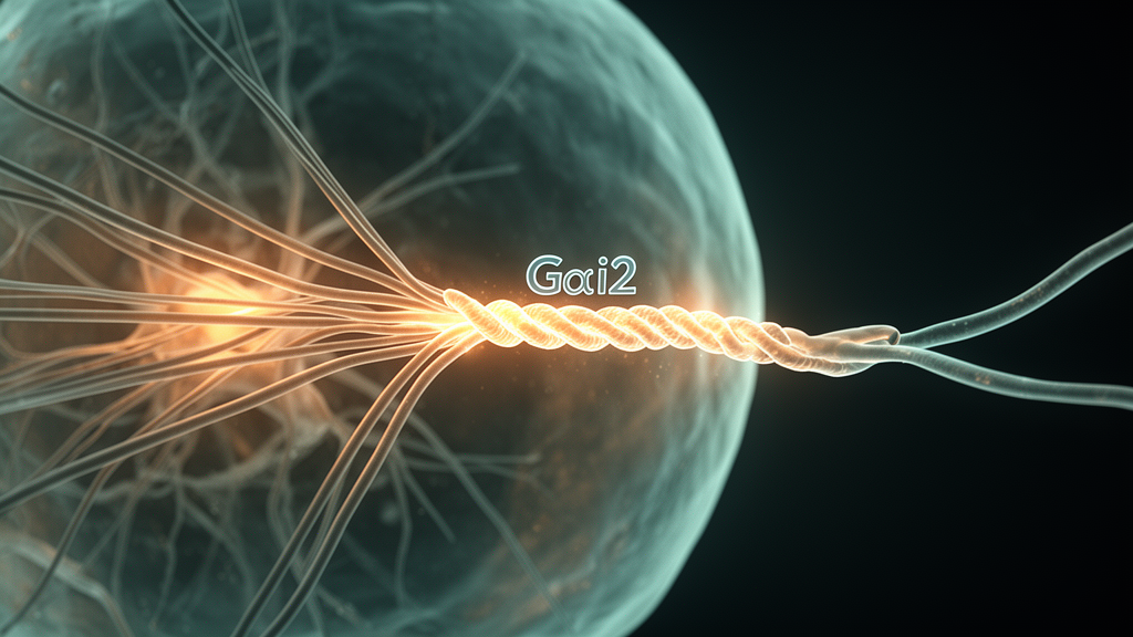
The identity of that other filamentous partner became clear when the researchers depolymerised microtubules with nocodazole. Treated neurons developed dense, coiled formations of Gαi2 in their cell bodies, resembling the collapsed intermediate filament networks described in cultured cells after microtubule disruption. Labelling for neurofilaments, the dominant neuronal intermediate filament, confirmed that Gαi2 coincided with neurofilaments in both treated and control tissue. In unmanipulated tissue, neurofilaments were concentrated towards the baso-ventral side of the cell throughout differentiation, and Gαi2 accumulations were confined to neurofilament-enriched regions, with coils of the two proteins associated with one another.
Live imaging filled in the dynamics. Neurons expressing a fluorescently tagged neurofilament protein, NFM-EGFP, showed that as axon outgrowth initiated, dense neurofilament accumulations at the baso-ventral region extended into the core of the nascent axon. Where coiled formations were visible, the filaments unfurled, rather like twine unravelling from a ball, as they extended into the growing projection. Meanwhile, timelapse imaging of the centrosome showed that it sat at the tip of the retracting apical process during delamination, was delivered to the cell body opposite the future axon site at initiation, and then moved towards the base of the extending axon. EB3 plus-tip tracking revealed microtubule growth trajectories emanating from the centrosomal region towards the nascent axon, indicating that the centrosome acts as a microtubule-organising centre and that microtubule orientation correlates with the axis of axon formation.
To probe the relationship between Gαi2 and the two filament systems directly, the team used in-situ proximity ligation assays and lattice structured illumination microscopy. These approaches showed Gαi2 decorating intertwining microtubule and neurofilament networks in the proximal axon, and, crucially, present at and around points where the two networks made contact. STED super-resolution imaging followed by colocalisation analysis found significantly higher overlap between Gαi2 and the neurofilament marker NF-H than between either Gαi2 and TUBB3 or NF-H and TUBB3, and flipping one image channel as a control collapsed the overlap, confirming the spatial relationships were genuine rather than artefacts of dense packing.
The functional test came from RNA interference. Knockdown of Gαi2, which reduced axonal Gαi2 fluorescence by roughly 40 percent, increased the proportion of pre-initiation and initiating cells relative to extending cells, suggesting disrupted axon formation. Proximity ligation assays revealed fewer microtubule–neurofilament interactions in Gαi2-depleted cells, both before initiation and in cells with axons. In pre-initiation cells, depletion led to a build-up of neurofilaments in the cell body that remained asymmetrically distributed towards the front, while stable microtubules became uniformly distributed rather than front-enriched. In initiating cells, neurofilament enrichment in the nascent protrusion was reduced, whereas microtubule distribution was unaffected. Notably, Gαi2 depletion did not change the speed or distance travelled by EB3 comets, implying that the protein does not alter microtubule growth dynamics in these neurons.
Live imaging of depleted cells showed the consequences for axon stability. Whereas most control cells initiated a single major protrusion over a 6.5-hour observation window, the majority of Gαi2-depleted cells initiated two or more, and the mean persistence of individual protrusions was significantly reduced. Neurofilament tracking showed that depleted cells accumulated a significantly smaller proportion of their total cellular neurofilaments within initiating protrusions at the timepoint preceding collapse, compared with control cells at equivalent stages, which progressively enriched neurofilaments in the axon as outgrowth proceeded. The authors propose that Gαi2 facilitates engagement of microtubules with neurofilaments, allowing neurofilaments accumulated at the cell front to be drawn into the axon and stabilise the growing protrusion; without it, neurofilament entry is impaired, protrusions retract, and repeated attempts at initiation produce multiple unstable projections.
The study is, according to the authors, the first to examine neurofilament behaviour during axon initiation in vivo, and it identifies a previously unreported localisation of a G protein to the intermediate filament network. The precise molecular mechanism remains open: Gαi2 might act directly as a linker between the two filament systems, or as part of a larger protein complex, and the authors note that further studies are needed to characterise its function at the molecular level. They also caution that directly testing neurofilament function in vivo is technically difficult because of the filaments’ exceptional stability and functional redundancy among their subunits. Since Gαi2 is highly expressed throughout the developing vertebrate nervous system and mutations in the human GNAI2 gene have been associated with intellectual disability and periventricular nodular heterotopia, the findings offer a possible mechanism by which disruption of this protein could hinder formation of neuronal circuitry, though the authors frame this as an avenue for future investigation rather than a demonstrated disease link.
Subject of Research: The role of the G protein Gαi2 in coupling neurofilaments to microtubules during axon formation in the embryonic chick spinal cord
Article Title: Gαi2 promotes microtubule-driven incorporation of neurofilaments to stabilise nascent axons in vivo
Article References: Higgs, V. E., Anwas, D., Astapenka, A., Toro-Tapia, G., & Das, R. M. (2026). Gαi2 promotes microtubule-driven incorporation of neurofilaments to stabilise nascent axons in vivo. EMBO Reports. https://doi.org/10.1038/s44319-026-00971-6
Image Credits: AI Generated
DOI: 10.1038/s44319-026-00971-6
Keywords: Gαi2, neurofilaments, microtubules, axon initiation, chick spinal cord, cytoskeleton, neuronal polarity, super-resolution microscopy, live imaging, RNA interference, neural development, EMBO Reports
News Source: Cassandra Pierce. (October 28, 2026). Gαi2 couples neurofilaments to microtubules to stabilise nascent axons in chick spinal cord. Scienmag.