Skeletal muscle has a striking ability to rebuild itself after injury, and a new review argues that the key to that repair may lie in molecules produced during exercise. Writing in Frontiers in Physiology, researchers from the Affiliated Hospital of Wuhan Sports University and Wuhan Sports University in China synthesize evidence that metabolites generated during physical activity, including lactate, alpha-ketoglutarate, acetyl-CoA, NAD+ and S-adenosylmethionine, are not simply waste products of energy metabolism. Instead, they act as substrates, cofactors and signaling molecules that directly influence epigenetic enzymes, converting the transient metabolic shifts of a workout into stable chromatin modifications and gene expression programs that guide the behavior of muscle satellite cells, the stem cells responsible for regeneration.
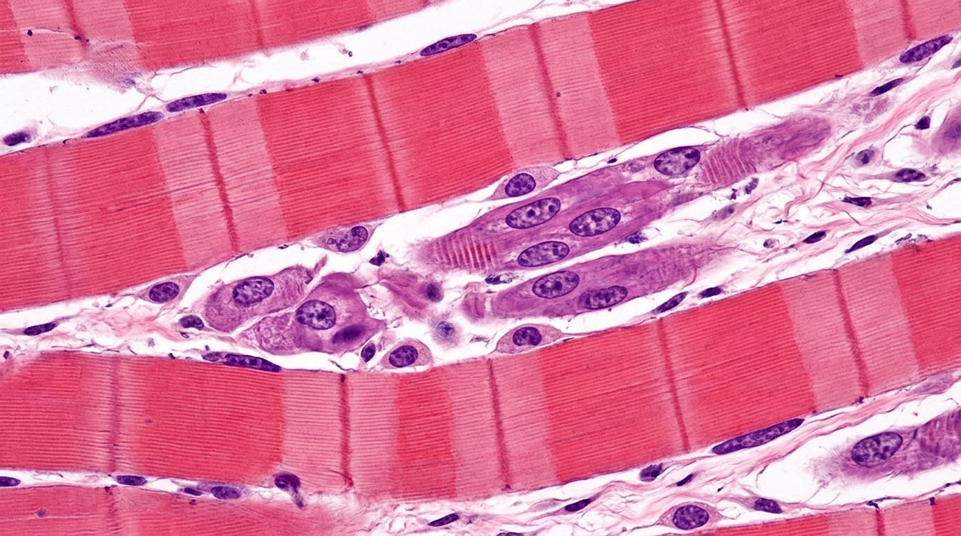
The review, published in the journal's Exercise Physiology section, centers on muscle satellite cells. Under physiological conditions these cells progress through a tightly choreographed sequence: activation, proliferation, differentiation from the resting state, and eventual fusion into mature muscle fibers. That progression is governed by the canonical myogenic regulatory cascade, marked by the sequential expression and functional coupling of Pax7, MyoD and myogenin. Aging, trauma and chronic diseases can impair satellite cell function, the authors note, leading to diminished regenerative efficiency, muscle atrophy, fibrosis and motor dysfunction, which is why understanding the molecular controls over cell fate matters for clinical intervention.
The authors define exercise as structured physical activity and divide it into two major modalities with distinct metabolic consequences. Endurance exercise involves prolonged, low-to-moderate intensity activity that relies predominantly on oxidative metabolism, whereas resistance exercise is characterized by high-intensity mechanical loading that promotes anabolic signaling and muscle hypertrophy. These modalities generate different metabolic environments and differentially regulate satellite cell behavior: endurance exercise primarily influences satellite cell differentiation and metabolic adaptation, while resistance exercise promotes activation, proliferation and myonuclear accretion, processes essential for muscle hypertrophy. The review also cites recent work from Kevin Murach and colleagues highlighting load-induced transcriptional and epigenetic programs in satellite cells, underscoring the importance of mechanical stimuli in shaping regenerative outcomes.
During regeneration, satellite cells undergo dynamic metabolic reprogramming tightly coupled to their fate transitions. Oxidative phosphorylation predominates in the quiescent state, but glycolysis is markedly upregulated during activation and proliferation to support rapid energy demand and biosynthesis, a shift the authors describe as resembling a Warburg-like effect. They stress that this reprogramming reflects a fundamental principle of cell growth rather than a pathological feature, citing the argument that cell growth is cell growth, and note that hypertrophying muscle fibers may partially reprogram their metabolism in a manner analogous to cancer cells while remaining tightly controlled and functionally beneficial. Following the proliferative phase, mitochondrial oxidative metabolism is progressively restored during differentiation as the cells mature.
Each metabolite carries a specific epigenetic assignment in the proposed framework. Increased glycolytic flux leads to lactate accumulation, which promotes histone lactylation, exemplified by the H3K18la mark deposited by acyltransferases such as p300, and facilitates gene expression associated with regeneration. Alpha-ketoglutarate, generated from the tricarboxylic acid cycle, serves as an essential cofactor for the JmjC-domain histone demethylases and TET-family DNA demethylases, helping remove inhibitory methylation marks such as H3K27me3 and H3K9me3 and thereby opening chromatin around myogenic genes like MyoD and myogenin. Acetyl-CoA acts as a substrate for histone acetylation, supplying acetyl groups to histone acetyltransferases including p300/CBP to create active marks such as H3K27ac and H3K9ac, enhancing chromatin accessibility and transcriptional activation. NAD+ regulates Sirtuin-mediated deacetylation, linking cellular redox status to transcriptional control, while S-adenosylmethionine functions as the primary methyl donor for DNA and histone methylation, influencing gene silencing and lineage commitment.
The review emphasizes that these metabolites do not operate in isolation but exist in competitive relationships that set the epigenetic state. Alpha-ketoglutarate competes with succinate and fumarate, which act as inhibitory metabolic signals that hinder alpha-ketoglutarate-dependent demethylation and establish a metabolic braking mechanism. Acetyl-CoA and NAD+ drive acetylation and deacetylation respectively, so their relative concentrations dictate whether chromatin is open or closed. The ratio of S-adenosylmethionine to its metabolite S-adenosylhomocysteine determines the efficiency of methyl transfer reactions and serves as an indicator of cellular methylation potential. Increased glycolysis also raises lactate levels and affects NAD+ regeneration, indirectly regulating deacetylation, so the metabolites collaboratively establish what the authors call a dynamic balance system of epigenetic regulation.
Timing matters as much as chemistry. In the early activation stage, an explosion of glycolytic flux produces rapid lactate accumulation, which the review says facilitates the transition of satellite cells from rest to activation through histone lactylation and by shaping the immune microenvironment. In the mid-phase proliferation stage, a dynamic equilibrium between acetyl-CoA and NAD+ governs chromatin accessibility and supports rapid cell expansion. In the late differentiation stage, mitochondrial oxidative metabolism prevails with increased levels of alpha-ketoglutarate and S-adenosylmethionine, which together erase inhibitory marks and maintain methylation homeostasis to promote the transformation of satellite cells into mature muscle fibers. Methylation changes during regeneration are described as highly dynamic and locus-specific: early activation is often associated with demethylation of myogenic regulatory genes, while later stages involve selective remethylation to stabilize transcriptional programs.
The two exercise modalities are proposed to shape different epigenetic trajectories. Endurance training is described as significantly activating the AMPK-ROS signaling pathway, characterized by an increased NAD+/NADH ratio and alpha-ketoglutarate accumulation, which enhances SIRT1-mediated deacetylation and JMJD3-mediated H3K27me3 demethylation, upregulates metabolic genes such as PGC-1alpha, promotes mitochondrial biogenesis and facilitates the phenotypic transformation of slow muscle fibers, effects considered particularly critical during later stages of satellite cell differentiation. Resistance training, by contrast, primarily activates the mTORC1-CaMKII signaling network, directing metabolic flow toward a high-throughput supply of acetyl-CoA and S-adenosylmethionine, producing elevated histone acetylation including H3K27ac and maintenance of DNA methylation homeostasis, and predominantly influencing the proliferation and fusion stages of satellite cells to promote hypertrophy and hypertrophic remodeling after regeneration.
Beyond immediate repair, the review addresses how repeated exercise may leave lasting marks. DNA methylation and histone modifications exhibit a degree of stability and can be inherited during cell division, potentially enabling satellite cells to respond more robustly to subsequent stimuli, a phenomenon the authors link to improved regenerative capacity after training. They cite evidence that DNA methylation patterns differ between myonuclear populations in skeletal muscle, revealing an epigenetic memory of prior adaptations, and that lifelong physical activity induces a satellite cell-dependent epigenetic fingerprint in muscle identity genes. Repeated fluctuations in metabolic status may progressively reshape the epigenetic landscape by continuously modulating S-adenosylmethionine and alpha-ketoglutarate levels, mediating long-term regenerative adaptation through these memory effects.
The clinical implications are framed cautiously. The authors propose that targeting the metabolic-epigenetic axis could offer new approaches for muscle injury, age-related sarcopenia and metabolic myopathy, through personalized exercise prescriptions, metabolic supplementation strategies such as NAD+ precursors, nutritional interventions to optimize methyl donor levels, and pharmacological regulation of energy metabolism and mitochondrial function. They also flag the limitations candidly: the causal relationship between metabolic changes and epigenetic regulation remains insufficiently defined, standardized protocols for intervention dosage and sequencing are lacking, individual differences in age, gender and basal metabolic status may affect efficacy, and current clinical evidence is limited, with most studies relying on animal models or in vitro experiments. Future work, they conclude, should integrate clinical trials with multi-omics analysis to develop stratified intervention models, advancing precision medicine for skeletal muscle regeneration.
Subject of Research: How exercise-induced metabolites regulate epigenetic enzymes controlling skeletal muscle regeneration
Article Title: Exercise-induced metabolites: messengers linking epigenetic enzymes and cell remodeling in skeletal muscle regeneration
Article References: Wang, W., Tian, Z., Bai, C., Li, P., Zuo, C., Ye, J., & Li, Z. (2026). Exercise-induced metabolites: messengers linking epigenetic enzymes and cell remodeling in skeletal muscle regeneration. Frontiers in Physiology, 17, Article 1885709. https://doi.org/10.3389/fphys.2026.1885709
Image Credits: AI Generated
DOI: 10.3389/fphys.2026.1885709
Keywords: skeletal muscle regeneration, muscle satellite cells, epigenetics, exercise metabolites, lactate, histone lactylation, acetyl-CoA, NAD+, S-adenosylmethionine, sarcopenia, muscle injury repair, metabolic-epigenetic axis
News Source: Juliet Wilcox. (November 5, 2026). Exercise metabolites act as messengers that reprogram gene activity during muscle repair. Scienmag.