
A groundbreaking advancement in neuroscience has emerged from a collaboration among researchers at Duke University School of Medicine, the University of Tennessee Health Science Center, and the University of Pittsburgh. These scientists have developed an unprecedented resource known as the Duke Mouse Brain Atlas. This innovative atlas promises to revolutionize the study of neurological disorders by offering a highly precise, three-dimensional map of the mouse brain, a model organism widely used in biomedical research. The atlas integrates microscopic-resolution images obtained through multiple imaging modalities, facilitating far greater accuracy in analyzing brain structure and pathology.
The Duke Mouse Brain Atlas represents the first truly stereotaxic and three-dimensional map of the entire mouse brain, distinguishing itself by capturing both the macro- and microscopic anatomical details. Unlike previous models that relied heavily on two-dimensional or partially reconstructed images, this atlas maintains the spatial integrity of brain structures as they exist in living animals. According to Dr. G. Allan Johnson, Charles E. Putman University Distinguished Professor of Radiology at Duke University, the stereotaxic nature of this atlas means it preserves critical external landmarks that guide experimental interventions in vivo, providing an accurate anatomical framework for scientific inquiry.
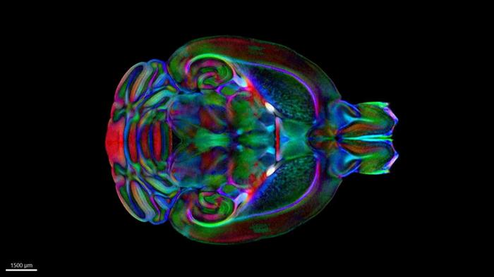
This stereotaxic precision addresses a longstanding challenge in neuroscience research: the discrepancy between high-resolution imaging techniques and their distortive effects on brain tissue. Traditional imaging, such as histological sectioning, offers exquisite cellular detail but sacrifices the anatomical context due to deformation during tissue processing. Conversely, non-invasive imaging like MRI can preserve spatial fidelity but struggles to resolve fine cellular features. The Duke Mouse Brain Atlas cleverly overcomes this by registering diverse data types—including magnetic resonance imaging (MRI), micro-computed tomography (microCT), and light sheet microscopy—into a unified coordinate system that combines both clarity and scope.
At the core of the atlas’s development is the application of diffusion tensor imaging (DTI), a sophisticated MRI technique, applied to postmortem mouse brains at an unparalleled resolution of 15 microns. This level of detail, achieved through decades of technological innovation at the Duke Center for In Vivo Microscopy, is approximately 2.4 million times higher than what typical clinical MRI scans can offer. Such resolution allows for an unprecedented visualization of neural pathways and microstructural brain components, enabling researchers to delineate subtle anatomical variations that are critical in diseases like Alzheimer’s and Huntington’s.
Enhancing the stereotaxic precision, the team incorporated microCT scans of the intact skulls to accurately localize bony landmarks. This step is essential because external cranial features serve as reference points during stereotaxic surgeries and imaging-based interventions. By aligning the internal brain imaging with these external landmarks, the atlas ensures compatibility with in vivo experimental setups, bridging the gap between ex vivo data and live animal procedures. Following this, light sheet microscopy was employed on extracted brains to reveal cellular and circuit-level architecture within the same spatial framework.
The integration of these imaging modalities into a single atlas represents a pinnacle of neuroimaging technology. By combining MRI’s strength in deep tissue penetration, microCT’s skeletal mapping ability, and the cellular resolution of light sheet microscopy, researchers created a comprehensive, multidimensional map that can be universally applied. This approach not only retains the natural anatomy but also standardizes data orientation and scale, a critical factor that enables cross-study comparisons and data sharing across laboratories globally.
Beyond its technical sophistication, the atlas is openly accessible and compatible with a variety of open-source visualization platforms, making it an invaluable educational and research tool. This democratization of advanced neuroimaging data means that even novice learners can appreciate the intricacies of brain anatomy, while seasoned neuroscientists gain a robust framework to quantify pathological changes with greater accuracy. Dr. Johnson highlights the potential impact on studies of neurodegenerative processes and toxic environmental exposures, where precise mapping is crucial to detect early and subtle cellular alterations.
In practical application, the Duke Mouse Brain Atlas is already serving as a vital tool in ongoing research. Investigators studying mouse models of Alzheimer’s disease are leveraging the atlas to track neurodegeneration longitudinally, correlating structural deterioration with functional deficits. Similarly, projects focused on Huntington’s disease and exposure-related neurotoxicity are utilizing this resource to unravel how diverse insults affect neural circuits at the micro- and macro-scale, paving the way for better therapeutic strategies.
The atlas also opens new avenues for multimodal neuroimaging studies, wherein data from behavioral assays, gene expression analyses, and electrophysiology can be anchored to a consistent anatomical framework. This compatibility will enhance integrative neuroscience approaches, facilitating discoveries that connect molecular, cellular, and systems-level phenomena. The standard space provided by the atlas reduces variability and enhances reproducibility, addressing significant challenges in preclinical neuroscience research.
In the broader context of animal models in neuroscience, the Duke Mouse Brain Atlas sets a new standard. It encourages the refinement of experimental designs by providing precise coordinates for targeting specific brain regions, optimizing stereotaxic interventions, and improving outcomes in functional and structural brain studies. This level of detail empowers researchers to discern subtle phenotypic differences and to better understand the complexities of brain organization and disease pathology in murine systems.
The atlas is slated for publication in the journal Science Advances on April 30, 2025, marking a significant milestone in neuroimaging research. The collaborative effort is bolstered by NIH funding through the National Institute of Neurological Disorders and Stroke and the National Institute on Aging, underlining the atlas’s importance in advancing neurological health research. The project’s leadership and technical expertise reflect decades of cumulative innovation, positioning the atlas as a foundational resource for the neuroscientific community.
The Duke Mouse Brain Atlas ultimately exemplifies how cutting-edge imaging technologies can be synthesized into a coherent and impactful research tool. It offers a detailed, spatially accurate, and accessible map of the mouse brain, empowering studies ranging from basic neuroscience to translational research in neurodegenerative diseases. As researchers continue to employ and refine this atlas, it promises to accelerate discoveries that could improve diagnostics, treatment, and our fundamental understanding of the brain.
—
Subject of Research: Animals
Article Title: The Duke Mouse Brain Atlas: MRI and light sheet microscopy stereotaxic atlas of the mouse brain
News Publication Date: 30-Apr-2025
Web References: http://dx.doi.org/10.1126/sciadv.adq8089
Image Credits: Duke University School of Medicine
Keywords: Neuroimaging, Magnetic resonance imaging, Alzheimer disease, In vivo imaging, Mouse models, Brain structure
Tags: advanced neuroscience toolsanatomical details in neurosciencebrain pathology studybrain structure analysiscollaboration in scientific researchDuke Mouse Brain Atlasexperimental interventions in vivomicroscopic-resolution imagingmouse model in biomedical researchneurological disorder researchstereotaxic brain atlasthree-dimensional brain mapping


