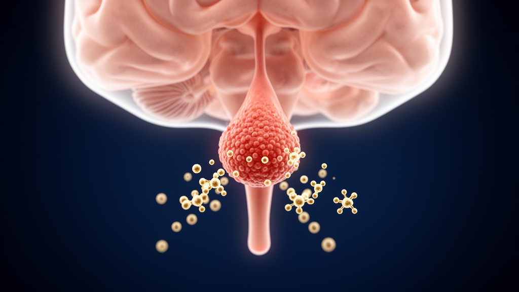
A single patient treated with the immune checkpoint inhibitor pembrolizumab has given researchers a rare window into one of the least expected complications of modern cancer immunotherapy: a collapse of the pituitary gland’s ability to produce the water-balancing hormone arginine vasopressin. The case, reported by physicians at the Cantonal Hospital Grisons in Chur, Switzerland, and published in Frontiers in Endocrinology, describes a 59-year-old man with metastatic lung adenocarcinoma who developed profound thirst, excessive urination, and dangerously elevated blood sodium only after receiving glucocorticoids for a separate immune-mediated colitis. The authors describe this timing as compatible with glucocorticoid-induced unmasking of vasopressin deficiency, while acknowledging alternative explanations such as fluid losses from colitis.
Immune checkpoint inhibitors such as pembrolizumab work by releasing the molecular brakes that tumors place on T cells. Pembrolizumab blocks programmed cell death protein 1, or PD-1, a receptor on immune cells whose engagement normally dampens T-cell activity and can drive T-cell destruction. By inhibiting this pathway, the drug enhances tumor regression, but the same immune liberation frequently triggers immune-related adverse events that can strike virtually any organ, most often the skin, gastrointestinal tract, and endocrine glands. Among endocrine complications, thyroid, pituitary, adrenal, and beta-cell dysfunction are the most common, in that order. Hypophysitis, inflammation of the pituitary, is seen most often with combined CTLA-4 and PD-1/PD-L1 regimens and least often with PD-1 inhibitor monotherapy, and men appear more frequently affected.
The patient arrived at the emergency department in 2024 with two weeks of diarrhea and vomiting and roughly five kilograms of weight loss. He had been receiving pembrolizumab, 200 mg intravenously, for twelve months, a total of nineteen cycles, with the most recent dose six days before admission. Stool cultures and tests for norovirus and Clostridium difficile were negative. A CT scan performed nine days earlier had shown no gastrointestinal pathology, but colonoscopy revealed erythematous changes of the colonic wall, and biopsies of the terminal ileum and colon confirmed drug-induced toxic colitis. A follow-up CT showed diffuse thickening of the rectum and sigmoid colon and a markedly distended stomach.
Because concurrent gastroparesis caused persistent vomiting, the team inserted a triluminal tube eleven days after admission to decompress the stomach and permit enteral nutrition. High-dose intravenous methylprednisolone, 80 mg daily for six days, was started, transitioning later to oral prednisolone with a taper. What followed was the diagnostic puzzle at the heart of the report. The patient developed marked thirst, initially attributed to his diarrhea and vomiting, and progressive polyuria, initially read as a consequence of increased fluid intake and substitution therapy. Nine days after admission, and three days after glucocorticoid initiation, he developed acute hypernatremia, with serum sodium climbing to 161 mmol/L against a reference range of 136 to 146.
Intravenous 5% glucose solution and free water delivered through the triluminal tube failed to bring the sodium down. The team began intranasal desmopressin, a vasopressin analogue, at 10 micrograms daily. The response was rapid: serum sodium fell to 144 mmol/L by day twelve, serum osmolality nearly normalized to 298 mmol/kg, and urine osmolality jumped from 132 to 512 mmol/L within a single day.
Supporting evidence came from further endocrine testing. Copeptin, a stable surrogate marker for vasopressin secretion, was undetectable, although the authors caution that the sample was drawn while serum sodium was already within the normal range, which limits its interpretive value. Thyroid function showed low-normal TSH with free triiodothyronine and free thyroxine below normal, suggesting possible central hypothyroidism, and levothyroxine was started. Follicle-stimulating hormone was low with normal luteinizing hormone and testosterone, so no gonadal replacement was given. ACTH was low as well, but secondary adrenal insufficiency could not be definitively confirmed because glucocorticoid therapy itself suppresses ACTH, and the same confounding applies to the thyroid results, since glucocorticoids also suppress TSH.
Brain MRI performed shortly after admission showed a normal sella turcica and pituitary gland, with no cerebral metastasis or pituitary enlargement. That, too, fits the expected pattern. In most patients with immune checkpoint inhibitor-induced hypophysitis, MRI shows pituitary enlargement and hypoenhancing lesions of the anterior lobe, but scans obtained within a month of diagnosis reveal substantially less pituitary swelling in patients treated with PD-1 inhibitors than in those receiving the CTLA-4 inhibitor ipilimumab. The authors also note an important limitation: their MRI was a standard brain protocol acquired to exclude metastasis, not a dedicated high-resolution sella study with thin cuts, so subtle posterior pituitary abnormalities, such as loss of the normal T1-hyperintense bright spot, could have been missed.
To place the case in context, the authors systematically searched PubMed and MEDLINE between April 2025 and April 2026 for case reports of pituitary dysfunction during pembrolizumab monotherapy, excluding combination regimens and cases without documented pituitary hormonal impairment. Thirty-two case reports met the criteria. Across these, pembrolizumab-triggered hypophysitis often affected multiple hormonal axes, with the adrenal axis most frequently impacted, while reports of arginine vasopressin deficiency remained rare. Only two comparable published cases of vasopressin deficiency with pembrolizumab monotherapy were identified, one of which also showed pituitary enlargement and stalk expansion on MRI while the other showed no infiltrative lesion. Five reports described secondary hypothyroidism, and reviewed cases typically presented with nonspecific symptoms such as anorexia and fatigue, mirroring this patient, though his symptoms could also be attributed to the malignancy and colitis.
The mechanistic explanation for the delayed unmasking centers on cortisol. Evidence reviewed by the authors suggests that cortisol deficiency compensates for absent antidiuretic hormone activity through several routes. Cortisol normally reduces the sensitivity of renal V2 receptors to vasopressin and limits aquaporin channel insertion into collecting duct cells, so its absence enhances whatever residual vasopressin activity exists. Corticotropin-releasing hormone stimulates both ACTH and vasopressin secretion, and without cortisol, negative feedback raises CRH and thereby vasopressin release. Cortisol also exerts mineralocorticoid-like effects promoting renal sodium reabsorption; its deficiency causes sodium and volume depletion that indirectly stimulates vasopressin secretion, and reduced glomerular filtration rate and tubular water excretion reinforce the same signal. When glucocorticoid replacement restored these pathways, the underlying vasopressin deficiency surfaced as hypernatremia and polyuria.
The authors are careful about the limits of their diagnosis. ACTH was measured only seven days after glucocorticoid initiation, making it impossible to distinguish secondary adrenal insufficiency from iatrogenic suppression, and no hypertonic saline infusion test was performed, so vasopressin deficiency cannot be substantiated by biochemistry alone. Colitis with substantial fluid losses offered an alternative explanation for the thirst and hypernatremia and delayed evaluation, and it remains possible that those losses contributed to the sodium rise. Still, the patient maintained normal serum sodium after hospitalization and after complete resolution of the colitis while continuing desmopressin, which the authors take as support for their hypothesis. His course was otherwise rocky: three colitis relapses during corticosteroid therapy, one complicated by hypovolemic shock and acute kidney injury requiring intensive care, an immediate allergic reaction to infliximab, and successful vedolizumab during the third relapse. At follow-up he remained stable on levothyroxine and desmopressin. The team concludes that because vasopressin deficiency in the setting of pembrolizumab-induced hypophysitis is rare and poorly investigated, clinicians should keep a low threshold for evaluating pituitary hormone deficiencies in immunotherapy patients with nonspecific symptoms, while noting that routine comprehensive pituitary screening of all asymptomatic patients on PD-1 monotherapy is not uniformly recommended.
Subject of Research: A rare case of arginine vasopressin deficiency unmasked by glucocorticoid therapy in pembrolizumab-induced hypophysitis
Article Title: Arginine vasopressin deficiency following pembrolizumab-induced hypophysitis: a case report and review of the literature
Article References: Dold, A. R., Jeker, R., & Mark, M. (2026). Arginine vasopressin deficiency following pembrolizumab-induced hypophysitis: a case report and review of the literature. Frontiers in Endocrinology, 17, Article 1976186. https://doi.org/10.3389/fendo.2026.1976186
Image Credits: AI Generated
DOI: 10.3389/fendo.2026.1976186
Keywords: pembrolizumab, hypophysitis, arginine vasopressin deficiency, central diabetes insipidus, immune checkpoint inhibitors, immune-related adverse events, desmopressin, pituitary, hypernatremia, glucocorticoids, endocrinology, case report
News Source: Ophelia Keating. (October 29, 2026). Diabetes insipidus unmasked after steroids for pembrolizumab hypophysitis. Scienmag.