The study, published in Breast Cancer Research, combined single-cell mass cytometry of matched blood and tumor samples, imaging mass cytometry of intact tissue, and activity-based probes that detect catalytically active enzymes in place, analyzing specimens from 66 patients. The central message is that these proteases are not uniformly distributed: they occupy specific cellular and spatial niches, and their abundance does not simply track with the surface markers clinicians currently use to select patients for targeted therapy.
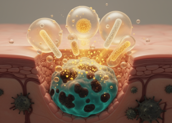
The work matters because cysteine cathepsins and legumain, an asparaginyl endopeptidase, are the molecular scissors that many antibody-drug conjugates rely on. If the relevant protease is scarce or inactive in a given tumor, the conjugate may deliver less drug than expected. Conversely, protease-rich microenvironments could amplify payload release. Understanding which cells carry which cathepsins, and where they sit relative to tumor cells, is therefore directly relevant to how such drugs are designed and prescribed.
The team first profiled the systemic immune landscape. Clustering of peripheral blood immune profiles separated patients into two groups, associated primarily with whether they had received neoadjuvant therapy and with tumor grade, and these clusters carried distinct patterns of cytokines and circulating protease levels. That systemic layer suggests the protease state of a patient is not confined to the tumor itself but is reflected in the bloodstream, raising the possibility — still to be tested prospectively — that blood-based readouts could eventually complement tissue biopsies.
Within tumors, single-cell analysis revealed pronounced interpatient heterogeneity in both tissue architecture and immune infiltration. Yet despite that variability, protease profiling uncovered reproducible modules tied to specific cell types.
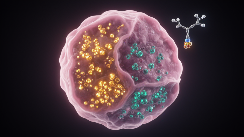
The cellular distribution differed sharply among family members. Cathepsins B and L were prominent in tumor-infiltrating immune cells and appeared variably in epithelial tumor cells. Cathepsin D showed a broader distribution across tumor cells, while cathepsin S remained more restricted. This division of labor means that a protease-cleavable drug optimized for one cathepsin may behave very differently depending on which immune and epithelial compartments in a given tumor express it.
Perhaps the most clinically provocative finding concerns HER2. In epithelial cells, HER2 expression did not consistently coincide with high cathepsin B or L abundance. That decoupling allowed the researchers to identify a limited subgroup of patients whose tumors combined HER2-high status with a protease-high state — precisely the combination most relevant to protease-cleavable antibody-drug conjugates targeting HER2. The implication is that target expression alone may be an incomplete criterion for patient selection: two HER2-positive tumors could differ substantially in their capacity to activate the same conjugate.
Spatial imaging added a geographic dimension to the map. Imaging mass cytometry localized cathepsins B and D to tumor-stroma interfaces and to macrophage-rich niches, the frontier zones where malignant cells meet immune and stromal compartments. Activity-based imaging mass cytometry then confirmed that catalytically active cathepsin B is genuinely present in human breast tumor tissue — not just the protein, but the functioning enzyme. That distinction matters because only active enzyme can cleave a drug linker.
The authors frame the work as a framework rather than a treatment advance: a protease-informed approach to patient stratification and to pairing biomarkers with protease profiles when designing targeted therapies. But by showing that HER2 abundance and proteolytic capacity can come apart, the findings argue that the next generation of antibody-drug conjugate strategies should read two properties of a tumor at once — what it displays on its surface, and what it can enzymatically unlock.
Subject of Research: Single-cell and spatial mapping of cysteine cathepsin proteases and their activity across human breast cancer ecosystems in relation to antibody-drug conjugate strategies
Article Title: Single-cell and spatial profiling of cysteine cathepsins identifies tumor states relevant to antibody-drug conjugates in breast cancer
Article References: Ćwilichowska-Puślecka, N., Małek, N., Gorzeń, O., Puślecki, T., Mlost, J., Nguyen, J., Dołęga-Kozierowski, B., Kasprzak, P., Sopel, M., Groborz, K., Szynglarewicz, B., Matkowski, R., & Poręba, M. (2026). Single-cell and spatial profiling of cysteine cathepsins identifies tumor states relevant to antibody-drug conjugates in breast cancer. Breast Cancer Research. https://doi.org/10.1186/s13058-026-02397-3
Image Credits: AI Generated
DOI: 10.1186/s13058-026-02397-3
Keywords: breast cancer, cysteine cathepsins, legumain, antibody-drug conjugates, mass cytometry, imaging mass cytometry, activity-based probes, HER2, tumor microenvironment, protease biomarkers, patient stratification, cystatins
News Source: Nathaniel Bowman. (October 29, 2026). Cysteine Cathepsins Mapped Across Breast Tumors Reveal States That May Shape Antibody-Drug Conjugate Use. Scienmag.