


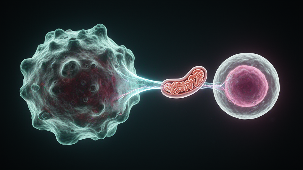
Mitochondria, the organelles that power nearly every cell in the body, are turning out to be far more mobile than biology textbooks ever suggested. A new review published in Genomics & Informatics on 13 January 2026 synthesizes a decade of evidence showing that intact mitochondria and their components travel between tumor cells, stromal cells and immune cells inside the tumor microenvironment. This intercellular trafficking, known as mitochondrial transfer, is emerging as a central mechanism by which cancers restore their energy supply, weaken antitumor immunity and resist chemotherapy, radiation and immunotherapy.
The routes of exchange are surprisingly varied. Tunneling nanotubes, thin F-actin-rich bridges that physically connect adjacent cells, allow direct transport of whole mitochondria and mitochondrial DNA. Extracellular vesicles such as exosomes and microvesicles carry mtDNA, mitochondrial proteins and even entire organelles between cells without any contact. More rarely, tumor cells fuse with mesenchymal stromal cells or macrophages, producing hybrid cells with shared cytoplasmic contents and enhanced oxidative phosphorylation and metastatic potential. Gap junctions involving connexin 43 may also facilitate vesicle-mediated delivery, and stressed cells can release mitochondrial fragments that neighbors internalize through endocytosis or phagocytosis. These routes are not mutually exclusive; a single tumor cell may exploit several in parallel, adapting to microenvironmental cues.
Molecular machinery governs this traffic. MIRO1, a mitochondrial Rho GTPase also known as RHOT1, links mitochondria to kinesin and dynein motor proteins through calcium-sensitive EF-hand motifs, enabling bidirectional movement along microtubules and actin filaments and stabilizing nanotube formation. DRP1 drives mitochondrial fission, dividing organelles into fragments small enough to travel through nanotubes, while MTFR2 promotes DRP1-dependent fission and increases the number of mitochondria available for transfer. Upstream, hypoxia stabilizes HIF-1α, which enhances transcription of MIRO1 and DRP1, and inflammatory cytokines activate Rho-family GTPases such as RAC1 and CDC42 that drive actin polymerization and filopodia extension. In hepatocellular carcinoma, hypoxic or nutrient-deprived stress upregulates HMGB1, RHOT1 and RAC1, promoting nanotube formation and restoring oxidative phosphorylation in neighboring tumor cells.
The metabolic consequences for tumors are dramatic. Tumor cells in nutrient-deprived or hypoxic niches often suffer mitochondrial dysfunction and reduced ATP production, and acquiring exogenous mitochondria replenishes functional electron transport chains. In glioblastoma models, astrocytes transfer mitochondria to tumor cells in a contact-dependent manner, and recipient cells show increased proliferation and self-renewal in vitro and in orthotopic mouse models, correlating with accelerated tumor growth and shortened host survival. In ovarian cancer, carcinoma-associated mesenchymal stromal cells donate mitochondria preferentially to mitochondria-poor tumor cells, rescuing respiration and proliferation and promoting metastatic spread in murine models. The flow can also reverse: tumor-to-fibroblast transfer reprograms stromal metabolism, induces cancer-associated fibroblast markers and stimulates release of pro-tumorigenic cytokines and extracellular matrix components.
Perhaps most striking is what this exchange does to the immune system. Tumor cells exploit tunneling nanotubes to hijack mitochondria from cytotoxic lymphocytes, particularly CD8-positive T cells and natural killer cells, depleting the immune cells’ respiratory reserves and weakening their cytotoxicity. Live cell imaging and electron microscopy have revealed cancer cells establishing actin-based intercellular tubes to siphon mitochondria directly from immune cells. Conversely, tumor cells can actively deposit dysfunctional mitochondria into infiltrating T cells. In human lung cancer and melanoma specimens, tumor-derived mitochondria were found inside tumor-infiltrating CD8-positive T cells, where they evaded autophagic clearance and caused accumulation of reactive oxygen species, driving the T cells into an exhausted state marked by elevated PD-1 and TOX and loss of effector cytokine production.
The clinical signal is notable. In melanoma and lung cancer patients, tumor-derived mitochondria carrying mtDNA mutations transferred to tumor-infiltrating lymphocytes produced metabolic dysfunction, senescence and poorer response to immune checkpoint inhibitors. The presence of tumor-derived mitochondrial DNA inside T cells correlated with poorer responses to anti-PD-1 immunotherapy. Similar impairments of cytotoxicity and cytokine production have been observed in NK cells and CD4-positive T cells receiving tumor-derived mitochondria. Tumor-associated macrophages add another layer: M2-polarized macrophages with fragmented mitochondria donated organelles to breast cancer cells that persisted as discrete ROS-generating units, promoting proliferation and invasiveness without necessarily boosting ATP production, indicating transfer can act as a signaling mechanism rather than a simple fuel delivery.
Not every transfer favors the tumor. A recent study found that osteocytes in bone donate mitochondria to metastatic cancer cells arriving in the bone niche, triggering the cGAS/STING innate immune pathway in those cancer cells and producing an antitumor immune response that suppressed growth of bone metastases. When this transfer was genetically blocked by deleting the mitochondrial fusion protein Mfn2 in osteocytes, cancer cells evaded immune detection and bone metastases progressed. The review’s authors stress that biological consequences of mitochondrial transfer depend critically on directionality, donor-recipient identity and the metabolic state of the transferred organelles, making it a context-dependent process rather than a universally tumor-promoting one.
Therapy resistance is another major theme. In acute myeloid leukemia, leukemic blasts under genotoxic stress stimulate bone marrow stromal cells to donate mitochondria through tunneling nanotubes, protecting the leukemia cells during chemotherapy; the transferred organelles raise oxidative phosphorylation and ATP levels, reducing dependence on glycolysis and conferring resistance to drugs such as cytarabine. Inhibiting the transfer process in AML increased leukemic cell apoptosis and improved survival in mouse models. In solid tumors, endothelial cells preferentially transfer mitochondria to breast cancer cells exposed to chemotherapeutic stress, dramatically enhancing resistance to doxorubicin. Mesenchymal stromal cells co-cultured with irradiated breast cancer cells transferred mitochondria that reduced radiation-induced ROS and improved survival, and horizontal transfer of mtDNA alone can restore respiratory function in tumor cells with radiation-damaged mitochondrial genomes, though direct in vivo evidence for radioresistance remains an active research area.
Tracing these events rigorously has required new tools. Dye-based approaches are prone to leakage, nonspecific uptake and confounding from cell fusion, so consensus guidance now emphasizes genetic labeling, dye-independent readouts and orthogonal validation. Genetically encoded reporters such as Mito-TRACER permanently label recipient cells that have taken up exogenous mitochondria, and in vivo lineage tracing with mitochondria-targeted fluorophores has confirmed host-derived signals inside implanted tumor cells. Endogenous mtDNA variants serve as natural barcodes: the MERCI pipeline analyzes mtDNA single-nucleotide variants within single-cell RNA sequencing datasets to reconstruct donor-recipient relationships in human tumors and has identified tumor-infiltrating T cells that acquired tumor-derived mitochondria. Complementary methods enrich mitochondrial variants at scale and pair mitochondrial genotypes with chromatin accessibility through mitochondrial single-cell ATAC-seq, enabling simultaneous lineage mapping and cell state readout.
Therapeutic strategies are still preclinical, and no approved therapies specifically inhibit intercellular mitochondrial transfer. Proposed approaches include blocking nanotube formation with actin polymerization inhibitors such as cytochalasin B or latrunculin, which decreased organelle transfer and increased chemotherapy sensitivity in co-culture experiments, and targeting CDC42 and other Rho-family GTPases, whose inhibition reduced mitochondrial exchange in vitro. Downregulating MIRO1 in donor cells can prevent efficient mitochondrial delivery, and GW4869, an inhibitor of exosome biogenesis, could reduce secretion of mtDNA-laden vesicles. In AML, pharmacological inhibition of NADPH oxidase 2 blocked stromal donation and sensitized leukemic cells to chemotherapy. On the immune side, early-phase studies combining metabolic modulators such as low-dose metformin with immune checkpoint inhibitors have shown potential to restore T-cell metabolism, and mitochondrial augmentation of cytotoxic T cells before adoptive transfer increased respiratory reserve and resilience against tumor-induced depletion. Safety remains a central concern, since nanotubes also support wound healing, neuronal recovery and immune communication in healthy tissue, and the authors caution that future therapies must selectively target tumor-specific mediators such as MIRO1 or TNFAIP2 while preserving physiological exchange elsewhere.
Subject of Research: Intercellular mitochondrial transfer between tumor, stromal and immune cells in cancer
Article Title: Mitochondrial transfer in cancer: mechanisms, immune evasion, and therapeutic opportunities
Article References: Ka, H. I., & Woo, H. G. (2026). Mitochondrial transfer in cancer: mechanisms, immune evasion, and therapeutic opportunities. Genomics & Informatics, 24(1), Article 2. https://doi.org/10.1186/s44342-025-00064-1
Image Credits: AI Generated
DOI: 10.1186/s44342-025-00064-1
Keywords: mitochondrial transfer, tunneling nanotubes, tumor microenvironment, immune evasion, T cell exhaustion, oxidative phosphorylation, therapy resistance, extracellular vesicles, mtDNA lineage tracing, MIRO1, DRP1, immunotherapy
News Source: Drew Townsend. (October 30, 2026). Cancer Cells Steal Mitochondria From Immune Cells to Survive and Evade Therapy. Scienmag.



