Cancer-associated fibroblasts (CAFs), the abundant stromal cells that populate solid tumors, are emerging as central architects of immunotherapy failure, and a new review argues that their metabolic rewiring is the key to understanding why. Published in Experimental Hematology & Oncology, the review synthesizes recent single-cell, spatial-omics and metabolomic studies showing that CAFs do not merely form passive scaffolding around tumors. Instead, they actively consume nutrients, export metabolites, remodel the extracellular matrix and reprogram immune cells, creating microenvironments in which immune checkpoint blockade (ICB) struggles to work.
ICB has transformed treatment for many solid tumors, but durable benefit remains limited to a subset of patients because of primary or acquired resistance. The review’s authors contend that this heterogeneity cannot be explained by tumor-intrinsic genetic alterations alone. The tumor microenvironment (TME), with its mixture of immune cells, blood vessels and stromal populations, shapes immune cell composition, functional states and spatial organization, and thereby influences clinical responses. Among stromal cells, CAFs stand out as one of the most abundant and phenotypically diverse populations across solid malignancies.
Far from being uniform, CAFs display remarkable heterogeneity in origin and function. Tissue-resident fibroblasts are considered the predominant source in many tumors, but epithelial cells can acquire CAF-like features through epithelial-to-mesenchymal transition, endothelial cells through endothelial-to-mesenchymal transition, and bone marrow-derived mesenchymal stem cells can differentiate into CAF-like cells after recruitment to tumor sites. In an inflammation-associated gastric cancer model, lineage tracing showed that a considerable proportion of CAFs originated from bone marrow-derived mesenchymal stem cells, and genetic lineage tracing in mice revealed that tumor-associated pericytes can undergo pericyte-to-fibroblast transition within the tumor stroma.
Single-cell RNA sequencing has further refined this picture, identifying recurring functional states. Myofibroblastic CAFs (myCAFs), driven largely by transforming growth factor-beta signaling, express contractile and extracellular matrix genes such as ACTA2, TAGLN and COL1A1, depositing collagen-rich matrix that stiffens tissue and can restrict immune-cell infiltration. Inflammatory CAFs (iCAFs), shaped by IL-1-alpha-driven NF-kappaB and JAK/STAT signaling, secrete cytokines and chemokines including IL-6, IL-8, CXCL1 and CXCL12 that modulate immune cell recruitment. Antigen-presenting CAFs (apCAFs) express MHC class II molecules but generally lack the co-stimulatory molecules needed for effective T-cell activation; in pancreatic ductal adenocarcinoma, mesothelial cell-derived apCAFs drive naive CD4-positive T cells toward antigen-specific regulatory T cell differentiation, supporting immunosuppression.
Crucially, these states are not fixed. In three-dimensional co-culture experiments, pancreatic stellate cells exposed to tumor organoids acquired an iCAF phenotype, while two-dimensional culture favored myCAF differentiation, and switching conditions reversed the states. Metabolic cues also drive remodeling: in pancreatic cancer, glutamine deprivation activates a MEK-ERK-dependent inflammatory program in myCAFs, whereas macropinocytosis, the scavenging of extracellular protein, preserves the myCAF state, and disrupting macropinocytosis promotes myCAF-to-iCAF conversion in vivo. The review stresses that CAF functions can be dual: in pancreatic cancer, FAP-positive CAFs are tumor-promoting while alpha-SMA-positive CAFs are tumor-restraining, so depleting the wrong subset can worsen outcomes.
The metabolic reprogramming itself is driven by multiple forces. Tumor-derived signals such as TGF-beta1 and platelet-derived growth factor can shift CAFs from oxidative phosphorylation toward aerobic glycolysis, partly through miR-424-dependent downregulation of the metabolic enzyme IDH3-alpha, which stabilizes HIF-1-alpha and promotes glycolytic gene expression. Cancer-cell-derived exosomes carrying miR-105 induce MYC-dependent metabolic reprogramming in stromal fibroblasts, and in triple-negative breast cancer, exosomes deliver integrin beta4 protein that triggers BNIP3L-dependent mitophagy and lactate production. The classic reverse Warburg effect, in which cancer cells induce aerobic glycolysis in neighboring fibroblasts that then export lactate and pyruvate to fuel tumor cell mitochondria, is reinforced by tumor-associated oxidative stress and hypoxia; in breast cancer, hypoxia activates ATM kinase in stromal fibroblasts, which phosphorylates the glucose transporter GLUT1 and increases lactate production.
Metabolite exchange, however, runs in both directions. In pancreatic cancer, highly glycolytic cancer cells consume most available glucose, leaving neighboring CAFs glucose-restricted; those CAFs take up cancer-derived lactate through the transporter MCT1 and burn it in the tricarboxylic acid cycle. In ovarian cancer, glutamine scarcity induces a glutamine-synthetic program in CAFs, whose high glutamine synthetase activity complements glutamine consumption by cancer cells. Epigenetic mechanisms add another layer: tumor-cell-derived lactate increases alpha-ketoglutarate in mesenchymal stem cells, activating TET enzymes during their differentiation into CAFs, while the enzyme NNMT consumes the methyl-donor S-adenosylmethionine in ovarian CAFs, reducing H3K27 trimethylation and driving stromal activation.
These metabolic programs translate directly into immune exclusion and dysfunction. In soft tissue sarcoma, CAFs at the tumor margin rely on GLUT1-dependent glycolysis to secrete CXCL16, impeding CD8-positive T-cell entry into the tumor core; pharmacological GLUT1 inhibition reduced CAF glycolytic activity, restored T-cell infiltration and enhanced chemotherapy sensitivity. FAP-positive CAFs overexpress the lncRNA LINC01711, boosting glycolytic flux and MCT4-dependent lactate export to form a metabolic barrier against T cells, and silencing LINC01711 restored antitumor immunity and sensitized tumors to anti-PD-1 therapy. In oral squamous cell carcinoma, TDO2-expressing CAFs secrete CXCL9, CXCL10 and CXCL11, recruiting T cells to peripheral stromal regions while limiting penetration into tumor nests.
Within immunosuppressive niches, CAFs disarm effector cells and empower suppressive populations. CAFs are a major non-immune source of the adenosine-generating enzyme CD73, whose expression is sustained by A2B receptor signaling in a feedback loop that suppresses CD8-positive T-cell function. Melanoma-associated fibroblasts deplete L-arginine through arginase, limiting mTOR-dependent protein synthesis and cytotoxic effector function in T cells. In pancreatic cancer, PLA2G2A-positive CAFs secrete an enzyme that suppresses MAPK/ERK and NF-kappaB signaling in infiltrating T cells, leaving them dysfunctional despite their presence, and CAF-expressed NNMT promotes CD8-positive T-cell suppression through EZH2-dependent epigenetic reprogramming. Natural killer cells are similarly targeted: in hepatocellular carcinoma, CAFs impair NK-cell cytotoxicity through prostaglandin E2 secretion and IDO-mediated tryptophan catabolism, while in gastric cancer, CAF-derived FSTL1 induces ferritinophagy in NK cells, increasing their susceptibility to ferroptosis. CAF-derived lactate in prostate cancer skews CD4-positive T cells away from Th1 differentiation and toward FoxP3-positive regulatory T cells.
Myeloid cells are equally reshaped. In lung cancer, lactate-activated DESMIN-positive CAFs secrete IL-8 that recruits tumor-associated macrophages and favors M2-like polarization, limiting ICB efficacy. In triple-negative breast cancer, CAFs promote monocyte differentiation toward lipid-accumulating macrophages, and an immunosuppressive STAB1-positive TREM2-high lipid-associated macrophage subset is enriched among patients resistant to ICB. A reciprocal macrophage-CAF circuit involving the nicotinamide metabolism enzymes NAMPT and NNMT modulates anti-PD-1 responsiveness, and in urothelial bladder cancer, CAF NNMT promoted macrophage infiltration and resistance to PD-L1 blockade, with pharmacological NNMT inhibition reducing macrophage infiltration and enhancing anti-PD-L1 efficacy in murine models. CAFs also drive neutrophil extracellular trap formation through secreted amyloid-beta, and NET abundance is associated with poor ICB response in non-small cell lung cancer.
Therapeutically, the field is learning that blunt approaches fail. FAP-targeted antibodies such as sibrotuzumab showed limited antitumor activity in a phase I trial, the FAP enzyme inhibitor talabostat produced no objective responses in metastatic colorectal cancer, and FAP-specific CAR T cells, though effective in mice, induced severe bone toxicity because FAP is expressed in normal mesenchymal cells. Matrix-directed strategies fared unevenly: the LOXL2 antibody simtuzumab failed to improve survival in metastatic pancreatic cancer, and the hyaluronidase PEGPH20 failed its phase III HALO-301 trial despite earlier biomarker-linked signals. Inhibiting CAF-activating pathways shows more promise: the TGF-beta receptor I inhibitor galunisertib prolonged overall survival with gemcitabine in a randomized phase II pancreatic cancer trial, and the NOX1/4 inhibitor setanaxib combined with pembrolizumab was associated with longer progression-free and overall survival and increased intratumoral CD8-positive T-cell infiltration in a phase II trial in head and neck squamous cell carcinoma.
The review’s authors are careful about what metabolic targeting can and cannot yet deliver. Current evidence does not show that it is broadly superior to conventional strategies, most metabolic interventions remain preclinical, and the same metabolic pathway may exert distinct or opposing functions across CAF subsets and disease stages. Many metabolic targets are shared with malignant cells, immune cells and normal tissues, raising specificity and toxicity concerns, and CAF metabolic plasticity means that inhibiting one pathway may simply trigger compensatory switching. The authors call for spatially and temporally resolved functional validation, combining spatial transcriptomics, spatial metabolomics, single-cell multi-omics and stable-isotope tracing with organoid and humanized models, to convert largely associative observations into clinically actionable strategies. CAF metabolic reprogramming, they conclude, should be viewed not as an isolated intracellular event but as an integral determinant of the tumor immune ecosystem and of whether immunotherapy ultimately succeeds.
Subject of Research: How metabolic reprogramming of cancer-associated fibroblasts shapes the tumor immune microenvironment and resistance to immune checkpoint blockade
Article Title: Metabolic reprogramming in cancer-associated fibroblasts: orchestrating the tumor immune microenvironment and response to immune checkpoint blockade
Article References: Gan, L., Liu, Z., Cao, L., Meng, X., Liu, X., Wang, X., & He, Y. (2026). Metabolic reprogramming in cancer-associated fibroblasts: orchestrating the tumor immune microenvironment and response to immune checkpoint blockade. Experimental Hematology & Oncology, 15(1), Article 104. https://doi.org/10.1186/s40164-026-00837-6
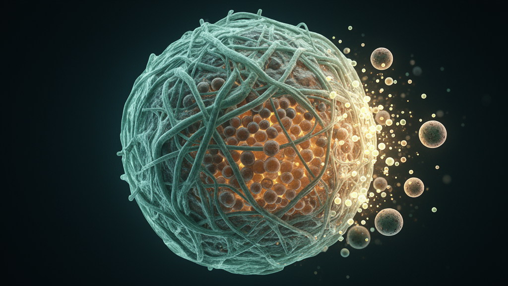
Image Credits: AI Generated
DOI: 10.1186/s40164-026-00837-6
Keywords: cancer-associated fibroblasts, metabolic reprogramming, tumor microenvironment, immune checkpoint blockade, immunotherapy resistance, single-cell omics, lactate, tumor-associated macrophages, regulatory T cells, extracellular matrix, TGF-beta, NNMT
News Source: Nathaniel Bowman. (October 28, 2026). Cancer-associated fibroblasts rewire tumor metabolism to block immunotherapy. Scienmag.