A Korean research team has developed a soft culture membrane intended to place intestinal organoid cells in a laboratory environment that more closely reproduces the biochemical composition and physical properties of the native intestine. The material combines an ultrathin nanofibrous scaffold with decellularized extracellular matrix derived from porcine colon tissue. When human colon organoid-derived epithelial cells were grown on the membrane, the researchers observed enhanced characteristics associated with intestinal stem-cell maintenance and proliferation, increased genetic markers associated with certain secretory cell lineages, and an epithelial layer with morphology more similar to native intestinal tissue. The peer-reviewed study was published online in Advanced Healthcare Materials.
The work addresses a mismatch between the cells used in intestinal models and the surfaces on which they are cultured. Intestinal organoids are miniature, stem cell-derived models that reproduce diverse cell types and functions of the native intestine. They are widely used to study intestinal diseases and evaluate drug responses. For some experiments, cells derived from these organoids are grown as a flat layer on a thin culture membrane. Although the cells may represent the intestine, the environment supporting them does not fully resemble it. Conventional culture platforms are made from synthetic materials that are much stiffer than native intestinal tissue and do not fully reproduce its tissue-specific extracellular matrix.
That difference can influence cellular behavior because epithelial cells respond to both biochemical and physical information. Proteins and growth factors are among the biochemical signals that can affect cells, while the stiffness and structural properties of the surrounding material provide physical cues. The release notes that such physical properties can influence cell proliferation, differentiation and tissue formation. The researchers therefore considered both the molecular composition and the physical behavior of the substrate when designing a more physiologically relevant intestinal culture system. Their objective was not simply to add selected soluble factors, but to combine tissue-derived biochemical components with a physical environment closer to native intestinal tissue.
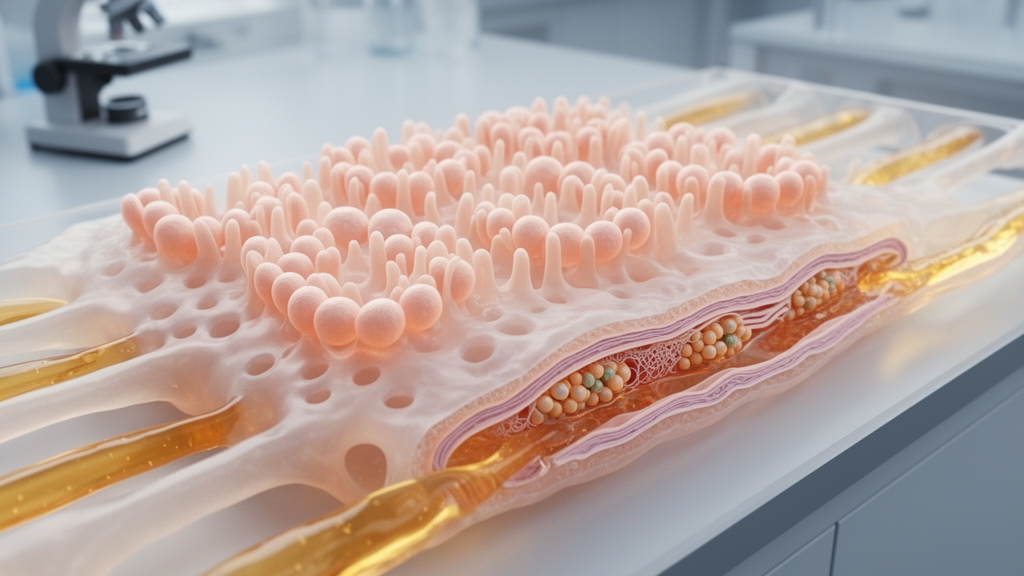
The team first used electrospinning to fabricate an ultrathin nanofibrous scaffold made from fibers far thinner than a human hair. It then incorporated decellularized extracellular matrix, or dECM, obtained from porcine colon tissue. Decellularization removes cellular components while preserving extracellular-matrix components. The resulting membrane, called a C-NaDE membrane, was designed to combine the structural stability of the nanofibrous scaffold with the tissue-specific biochemical components of colon-derived dECM. Unlike conventional synthetic culture membranes, it also provided a softer physical environment, allowing cells to be cultured under conditions that more closely approximated their physiological surroundings.
In laboratory testing, the researchers cultured human colon organoid-derived epithelial cells on the new membrane. The cells exhibited enhanced characteristics associated with intestinal stem-cell maintenance and cell proliferation. The team also observed increases in genetic markers associated with certain secretory cell lineages, which the release describes as relatively difficult to detect under conventional culture conditions. In addition, the epithelial layer displayed morphological characteristics more closely resembling those of native intestinal tissue. These observations concern cells in laboratory culture; they do not constitute evidence of a treatment effect or a clinical benefit in people.
The study’s key advance is the integration of two classes of environmental cues in a single membrane. Conventional approaches have often focused on supplementing individual proteins or growth factors, whereas the C-NaDE membrane combines biochemical material derived from intestinal tissue with a softer physical environment. In this way, the researchers attempted to reproduce both the composition and the physical character of the environment in which intestinal cells naturally reside. Their results indicate that the combined substrate affected several measured characteristics of the cultured epithelial cells, rather than being limited to one marker or a single cellular behavior.
The membrane can be incorporated into conventional cell-culture systems, and the researchers also propose its use in microfluidic organ-on-a-chip platforms. The release identifies intestinal disease modeling, drug-response testing, intestinal-barrier assessment and studies of interactions between the intestinal epithelium and gut microbiota as possible future applications. These are proposed uses rather than outcomes already established in the reported experiment. The current evidence specifically concerns human colon organoid-derived epithelial cells cultured on the laboratory membrane, so broader performance would require additional evaluation under the conditions needed for each proposed application.
Professor Dong Sung Kim of POSTECH’s Department of Mechanical Engineering and Graduate School of Convergence Science and Technology said the significance of the study lies in integrating tissue-specific intestinal components and a physiologically relevant physical environment within one culture membrane. He noted that organoid models reproduce diverse intestinal cell types, but monolayers typically rely on synthetic substrates whose biochemical and physical properties differ substantially from those of native tissue. Professor Jaeseung Youn of CHA University School of Medicine, the first author, said cells respond not only to biochemical factors such as growth factors and proteins but also to physical properties including stiffness. According to Youn, recreating the biochemical and biophysical microenvironment provides a foundation for intestinal models that more closely resemble the in vivo environment.
For researchers, the most immediate implication is that the material surrounding cultured cells can be treated as part of the model rather than as an inert support. The C-NaDE membrane links an electrospun nanofibrous structure with colon-derived dECM and provides a softer substrate for a flat intestinal epithelial layer. In the reported cell-culture experiment, that approach was associated with enhanced stem-cell maintenance and proliferation characteristics, increased markers for certain secretory lineages, and more native-like epithelial morphology. The platform may help laboratories investigate how intestinal epithelial cells respond when biochemical and biophysical cues are presented together, while its value for disease modeling, drug testing, barrier studies and microbiota research remains a direction for further development.
Subject of Research: Biomimetic culture membrane for human intestinal organoid epithelial cells
Article Title: Beyond plastic: A new culture membrane mimics the native intestinal environment
Article References: Beyond plastic: A new culture membrane mimics the native intestinal environment. Original publication (Bibliographic details unavailable.)
Image Credits: AI Generated
DOI: Not provided
Keywords: intestinal organoids, biomimetic membrane, decellularized extracellular matrix, nanofibrous scaffold, intestinal epithelium, stem-cell maintenance, cell proliferation, secretory cell markers, organ-on-a-chip, tissue engineering, Beyond, plastic
News Source: Gregory Coleman. (October 26, 2026). Biomimetic membrane recreates native-like conditions for intestinal organoid cells. Scienmag.