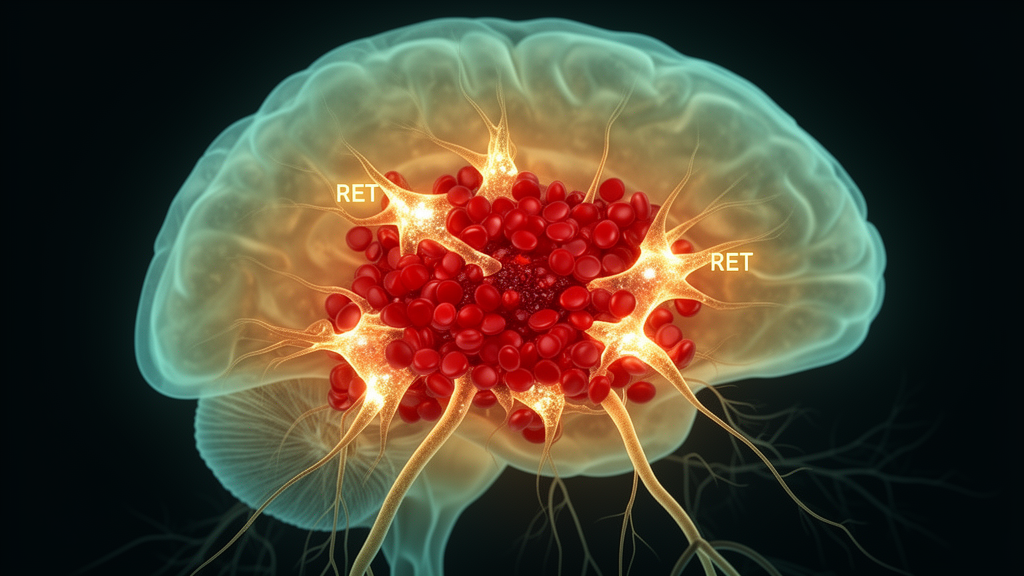
When a blood vessel ruptures inside the brain, the damage does not stop at the rim of the clot. Neurons surrounding the hematoma are highly vulnerable to the toxic aftermath of the bleed, yet they remain potentially salvageable, and understanding what decides their fate is a central goal of stroke research. A new study published in BMC Molecular and Cell Biology points to an unexpected player in that decision: RET, a receptor protein whose signaling appears to help astrocytes, the brain’s star-shaped support cells, shield injured neurons from programmed cell death.
The work builds on the research team’s earlier gene chip analysis, which suggested that an ASCL1-RET signaling axis is involved in neural injury after intracerebral hemorrhage. To follow up, the researchers set out to determine what RET actually does in the perihematomal region, the tissue immediately surrounding the bleed, and by what mechanism it might act. They established an in vivo mouse model of intracerebral hemorrhage alongside in vitro cultures of primary astrocytes and neurons.
Using Western blotting and immunofluorescence, the team tracked both total RET and its phosphorylated, activated form. Phosphorylated RET was significantly upregulated in the perihematomal region after hemorrhage, reaching its peak at day one. The signal was not confined to a single cell type: it co-localized with neurons, astrocytes, and microglia, indicating that RET activation is a broad early response in the injured neighborhood rather than a feature of one cell population alone.
To probe the protein’s function directly, the researchers turned to cell culture. When primary astrocytes were stimulated with hemin, a heme-derived compound used to mimic the blood exposure that follows hemorrhage, the astrocytes became reactive and RET phosphorylation increased. Critically, the conditioned medium collected from these hemin-stimulated astrocytes reduced neuronal apoptosis, an effect that disappeared when RET was silenced in the astrocytes using shRNA lentivirus. In other words, RET in astrocytes was required for the protective influence those cells exerted on neighboring neurons.
Neuronal cell death was assessed with a TUNEL assay and by measuring cleaved caspase-3 expression, two standard markers of apoptosis. Changes in PI3K/AKT phosphorylation were associated with the RET-dependent, astrocyte-mediated neuroprotection, suggesting that the PI3K/AKT pathway is part of the mechanism linking astrocytic RET signaling to neuronal survival. The authors conclude that astrocytic RET protects neurons from hemin-induced apoptosis and supports RET-dependent crosstalk between astrocytes and neurons after hemorrhage.
The findings are framed by the authors as supporting RET as a potential therapeutic target for neuroprotection after intracerebral hemorrhage. It is worth stressing what the study does and does not show. The evidence comes from a mouse model and from primary astrocyte and neuronal cultures, not from human patients, and the reported effects concern associations with PI3K/AKT phosphorylation rather than a demonstrated causal chain in vivo. No therapeutic intervention was tested in the study itself; the suggestion of RET as a target is a direction for future work rather than a validated treatment. The article was published as an open-access paper on 30 September 2026 and is currently available in a version that remains subject to further editorial processing before the final version of record replaces it.
Even with those caveats, the study adds a specific molecular thread to a growing picture of astrocytes as active participants in brain injury responses rather than passive bystanders. If RET-dependent astrocyte signaling proves robust in further preclinical work, it could offer a route to reinforcing the brain’s own protective circuits in the critical hours and days after a hemorrhagic stroke, when neurons around the hematoma are still salvageable.
Subject of Research: The role of the RET protein in astrocyte-mediated neuroprotection in the perihematomal region after intracerebral hemorrhage.
Article Title: The role of the RET protein in the perihematomal region after intracerebral hemorrhage
Article References: Geng, W., Song, Y., Ke, K., Shen, J., & Fang, Q. (2026). The role of the RET protein in the perihematomal region after intracerebral hemorrhage. BMC Molecular and Cell Biology. https://doi.org/10.1186/s12860-026-00606-7
Image Credits: AI Generated
DOI: 10.1186/s12860-026-00606-7
Keywords: RET protein, intracerebral hemorrhage, astrocytes, neuroprotection, neuronal apoptosis, PI3K/AKT signaling, perihematomal region, mouse model, glia-neuron crosstalk, ASCL1-RET axis, role, protein
News Source: Cassandra Pierce. (October 28, 2026). Astrocytic RET protein shields neurons after brain hemorrhage in mouse study. Scienmag.