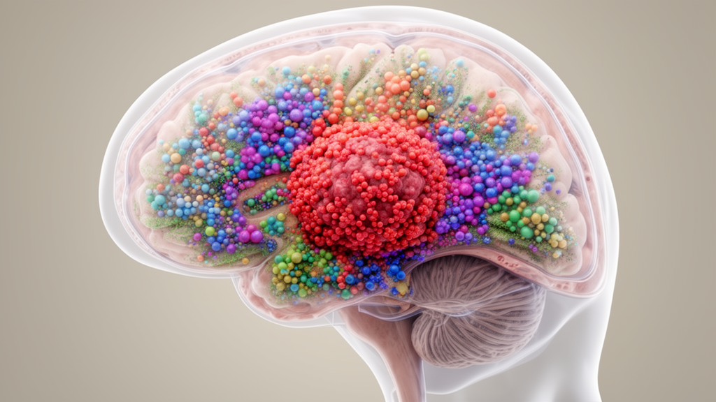
Glioblastoma, the most aggressive primary brain tumor in adults, has long frustrated researchers with its ability to evade treatment through sheer biological complexity. Now, a team of neurosurgeons and computational biologists from institutions across China has delivered one of the most methodologically rigorous portraits yet of the tumor’s inner ecosystem, combining machine learning, single-cell genomics, bulk RNA sequencing, and laboratory validation into a single reproducible framework. Their findings, published in Frontiers in Cell and Developmental Biology, do not claim to have discovered a new subtype of glioblastoma. Instead, they do something arguably more valuable: they show, with unusual statistical care, which parts of the tumor’s transcriptional landscape can be trusted, which associations are merely descriptive, and which popular narratives about immune-tumor crosstalk remain unproven.
The study reanalyzed the landmark GSE131928/SCP393 dataset originally generated by Neftel and colleagues, comprising 24,131 single cells from IDH-wildtype glioblastoma profiled across two sequencing platforms: Smart-seq2, which contributed 7,930 cells across 28 tumor labels, and 10x Genomics, which contributed 16,201 cells across 9 tumor labels. Rather than treating these platforms as interchangeable, the researchers audited every expression matrix on its verified scale, transforming the 10x data from TPM-like linear values to a log2(TPM/10 + 1) scale to match the deposited Smart-seq2 format. This meticulous attention to data provenance matters because cross-platform integration is notoriously fragile, and small errors in normalization can produce spurious biological conclusions that ripple through an entire field.
At the heart of the analysis lies the now-canonical framework of malignant cell states in glioblastoma. Rather than existing as a single fixed subtype, glioblastoma cells occupy four recurrent transcriptional programs: astrocyte-like (AC-like), mesenchymal-like (MES-like), neural-progenitor-like (NPC-like), and oligodendrocyte-progenitor-like (OPC-like). These states coexist within individual tumors and shift dynamically, reflecting the remarkable plasticity that makes the disease so difficult to treat. The research team recomputed these four broad states from the official module definitions, carefully excluding 287 records whose state modules had been corrupted by gene aliasing, leaving 6,576 malignant cells with valid state scores as the denominator for all downstream analyses. It is the kind of unglamorous data hygiene that rarely makes headlines but fundamentally determines whether a computational result can be reproduced by other laboratories.
To bridge the two sequencing platforms, the researchers built a machine-learning label-transfer model using a grouped five-fold logistic regression in which entire tumor labels were held out as groups during training. This design prevents information leakage between tumors, a common pitfall in single-cell machine learning. The model achieved an out-of-fold accuracy of 0.998 with a macro F1 score of approximately 0.985, and only 141 cells, or 0.87 percent, remained unassigned. When the researchers compared malignant-state fractions between the Smart-seq2 and 10x arms across the six tumor labels shared by both platforms, they found Spearman correlations ranging from 0.66 to 0.83, indicating moderate-to-strong concordance. All 11,448 shared-label malignant pseudobulk gene pairs tracked closely along the diagonal, confirming that the two platforms tell a coherent story about the same biological reality.
But the study’s most consequential finding may be what it did not find. A persistent narrative in glioblastoma research holds that macrophages infiltrating the tumor drive malignant cells toward a mesenchymal-like state, creating a self-reinforcing loop of immune suppression and therapeutic resistance. When the researchers directly tested this hypothesis at the tumor-label level, the result was a null: the MES-like fraction showed no significant association with macrophage fraction, yielding a Spearman rho of just 0.171 with a P value of 0.395 across 27 tumor labels. An adult-only sensitivity analysis produced a similarly negligible result. This null finding does not disprove macrophage-tumor interactions, but it does sound a cautionary note for a field that has sometimes promoted bulk-level correlations into directional mechanisms without adequate evidence.
Extending the analysis to bulk transcriptomics, the team turned to the TCGA-GBM cohort, reducing 176 expression aliquots through 158 primary tumors and 143 primary IDH-wildtype libraries to 142 unique patients. Using fixed gene signatures with coverage between 0.91 and 1.00, they computed patient-level state scores and found that the MES score correlated strongly with both immune (Spearman 0.547, P < 0.001) and stromal (0.552, P < 0.001) ESTIMATE scores, while the myeloid program showed an even more striking correlation with immune signatures at 0.954. Conversely, NPC and OPC scores were inversely associated with immune and stromal estimates. These patterns are biologically plausible, but the researchers are careful to note that bulk RNA cannot resolve which cell type produces which transcript, and that tumor purity and compositional variation may partly drive the observed associations.
A score-defined differential expression contrast between patients with high versus low MES and immune program scores yielded 8,620 genes below a false discovery rate of 0.05, with FCGR2A showing the strongest effect at a log2 fold change of 1.417 and a Hedges g of 2.041. Hallmark pathway analysis led by Interferon Gamma Response (normalized enrichment score 3.990, FDR q = 0.001) confirmed the immune character of the high-score group. Yet the researchers explicitly label these analyses as descriptive rather than independent discovery, because the grouping variable is derived from the very expression programs under investigation. This kind of intellectual honesty, distinguishing score geometry from genuine biological discovery, is increasingly rare and increasingly necessary in the era of large-scale computational oncology.
Survival and prediction analyses further tempered any temptation toward clinical overreach. A fixed median split of the combined MES/immune score produced median overall survival of 12.0 versus 14.0 months with a log-rank P value of 0.663, far from statistical significance. Cox model estimates shifted depending on covariate specification, with the hazard ratio for the MES/immune score moving from 1.095 (P = 0.314) in univariable analysis to 1.372 (95% CI 1.019–1.848, P = 0.037) only after adding Karnofsky performance status, a change the authors attribute to specification sensitivity and reduced sample size. The best nested cross-validated model for predicting 12-month mortality, a combined elastic net incorporating clinical and signature features, achieved an out-of-fold ROC-AUC of just 0.587, with permutation importance led by age and the myeloid program. These are workflow benchmarks, not deployable biomarkers.
To ground the computational findings in experimental data, the researchers performed RT-qPCR on four glioma cell lines (U87MG, LN229, U251, and T98G) alongside normal human astrocytes as a non-malignant reference. Four genes representing malignant, mesenchymal/myeloid, and angiogenic programs were all significantly overexpressed in glioma cells relative to normal astrocytes. CHI3L1 showed the most dramatic elevation, reaching up to 12.8-fold in U87MG cells, while SPP1 and VEGFA were upregulated 6- to 9-fold and 5- to 8-fold respectively, and STAT3 showed consistent 4- to 6-fold upregulation. These results align with prior literature implicating STAT3 in tumor signaling, AXL-linked therapy resistance within mesenchymal programs, and SPP1-mediated macrophage-tumor crosstalk, though the authors emphasize that cell-line expression data cannot validate a tumor-associated macrophage-derived mechanism in living tissue.
The study’s ultimate contribution lies not in a single dramatic discovery but in the framework it establishes. By demonstrating that established malignant cell states can be recovered reproducibly across sequencing platforms, that immune-associated expression programs covary with microenvironmental signatures in bulk tissue, and that popular mechanistic narratives require perturbational and spatial evidence before they can be accepted as causal, the researchers have provided a template for how integrated single-cell and bulk transcriptomics should be conducted in the era of reproducible computational biology. Independent cohorts, spatial profiling, paired longitudinal specimens, and functional perturbation experiments will be needed to convert these descriptive associations into actionable biology, but the foundation is now considerably more solid than it was before this analysis began.
Subject of Research: Machine-learning integration of single-cell and bulk transcriptomics to characterize glioblastoma malignant cell states and immune-associated tumor ecosystem programs
Article Title: Machine-learning-assisted integration of single-cell and bulk transcriptomics characterizes glioblastoma ecosystem states and immune-associated programs
Article References: Cao, J., Cheng, H., Wang, Y., Zhao, B., Wang, Y., & Jiang, X. (2026). Machine-learning-assisted integration of single-cell and bulk transcriptomics characterizes glioblastoma ecosystem states and immune-associated programs. Frontiers in Cell and Developmental Biology, 14, Article 1967483. https://doi.org/10.3389/fcell.2026.1967483
Image Credits: AI Generated
DOI: 10.3389/fcell.2026.1967483
Keywords: glioblastoma, single-cell RNA sequencing, machine learning, tumor microenvironment, macrophages, mesenchymal state, TCGA, RT-qPCR, label transfer, immune programs, transcriptomics, IDH-wildtype
News Source: Nathaniel Bowman. (October 16, 2026). AI-Powered Map of Glioblastoma Reveals Tumor Ecosystem States and Immune Programs. Scienmag.