In a pioneering advancement at the intersection of oncology and artificial intelligence, researchers from the ECOG-ACRIN Cancer Research Group have harnessed cutting-edge AI-driven methodologies to detect tertiary lymphoid structures (TLS) within thousands of high-resolution digital melanoma tumor images. This breakthrough significantly refines the identification of TLS—a vital biomarker linked to improved prognosis in operable stage III and IV melanoma patients—offering unprecedented accuracy and consistency compared to traditional pathological techniques, which are often laborious and prone to variability.
Tertiary lymphoid structures represent specialized immune cell aggregates that develop ectopically within tumor microenvironments. These formations, comprising T cells, B cells, and dendritic cells, emerge in response to chronic inflammation or neoplastic progression. TLS have been strongly correlated with enhanced immune infiltration and favorable patient outcomes across multiple cancer types, yet their integration into routine pathology workflows remains limited due to detection challenges. The ECOG-ACRIN researchers’ AI-enhanced approach seeks to surmount these hurdles by automating TLS detection through sophisticated image analysis.
The team’s investigation retrospectively analyzed an extensive cohort of 376 patients diagnosed with advanced, high-risk melanoma. By integrating digitized hematoxylin and eosin (H&E)-stained histologic slides with corresponding RNA sequencing datasets, the researchers established a definitive link between TLS presence and markedly improved overall survival. Derived from participants in the landmark E1609 clinical trial—which evaluated immune checkpoint inhibitors and cytokine therapies—this study leverages a robust dataset, positioning it to inform future prognostication and therapeutic stratification efforts.
Quantitative analysis within this cohort revealed TLS in approximately 55% of cases, with significant survival benefits observed in patients harboring TLS compared to those without. Specifically, five-year overall survival rates were 36.23% in TLS-positive patients, contrasting with 29.59% in the TLS-negative group. Intriguingly, patients exhibiting multiple TLS demonstrated an even greater survival advantage, underscoring the prognostic relevance of TLS density alongside presence. Additional stratification highlighted survival variability based on established clinical parameters such as AJCC tumor stage, patient age, sex, therapeutic modality, and tumor ulceration status.
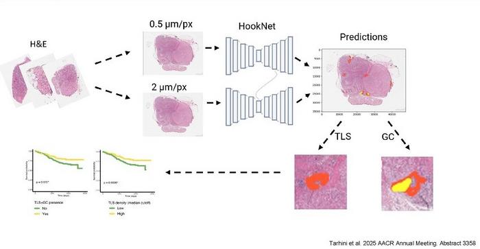
Central to these advancements is the deployment of HookNet-TLS, an innovative open-source deep learning algorithm explicitly designed for automated detection of TLS and germinal centers within digitized histological images. Originally developed for bioimage analysis, HookNet leverages convolutional neural network architectures to perform end-to-end segmentation and classification of complex tissue structures at high resolution. After initial application demonstrated promising outcomes, the researchers undertook model refinement to enhance predictive accuracy, thereby enabling robust quantification of TLS and associated germinal centers.
The implications of integrating such AI-driven tools into routine clinical workflows are profound. By automating the evaluation of TLS using low-cost, widely accessible H&E-stained samples, this approach promises to standardize assessments that have previously been subjective and resource-intensive. Moreover, the enhanced sensitivity and specificity in TLS detection could enable more accurate prognostication within the AJCC staging framework, ultimately informing personalized immunotherapy decisions and improving clinical outcomes for high-risk melanoma patients.
This research initiative, supported by funding from the National Cancer Institute, exemplifies the transformative potential of synergizing biomedical imaging, machine learning, and molecular oncology. The ability to rapidly and reproducibly quantify critical immune microenvironment components paves the way for integrating biomarkers like TLS into established diagnostic paradigms and therapeutic decision-making algorithms.
As highlighted by Dr. Ahmad A. Tarhini, lead investigator and professor at the Moffitt Cancer Center, “Our work showcases how openly accessible AI tools can revolutionize the prediction of survival and immunotherapy response by facilitating detailed immune structure analysis—ushering in a new era of precision oncology.” Co-investigator Dr. Xuefeng Wang emphasized the promise of foundation models like Gigapth in refining such analyses, pointing to ongoing developments that will enhance the robustness and applicability of these methods in broader clinical contexts.
The ability to detect TLS efficiently and accurately could reshape clinical conversations between physicians and patients, particularly regarding the potential benefits of immunotherapy in melanoma. As these AI methodologies mature, they hold promise not just for oncology but also for other immune-related diseases where tertiary lymphoid structures may play pivotal roles.
Tertiary lymphoid structures, by virtue of their composition and spatial organization, represent dynamic hubs of antitumor immune activity. Their detection and quantification have historically required expert pathologists to identify subtle histological features—a challenge complicated by interobserver variability and resource constraints. The successful deployment of AI algorithms like HookNet-TLS, which automate these tasks with high precision, addresses critical gaps in workflow efficiency and diagnostic standardization.
Furthermore, the public release of HookNet’s source code on platforms such as Grand Challenge fosters transparency and collaboration across the biomedical imaging and AI communities. This open-source ethos accelerates innovation, enabling researchers worldwide to adapt and refine algorithms for localized datasets, diverse cancer types, and extended biomedical applications.
The advancements demonstrated in this work are poised to be presented at the upcoming American Association for Cancer Research 2025 Annual Meeting in Chicago, where further insights into model performance and clinical applicability will be shared. The anticipated dissemination of these results will likely catalyze interest and investment in AI-facilitated pathology, heralding a paradigm shift in how immune biomarkers inform oncologic care.
By leveraging sophisticated AI frameworks to harness existing digital pathology resources, the ECOG-ACRIN team has unlocked new dimensions in melanoma prognostication, showcasing a scalable path forward for integrating machine learning into precision medicine.
Subject of Research: Artificial intelligence-driven detection of tertiary lymphoid structures in advanced melanoma for improved survival prediction
Article Title: Not explicitly provided in the content
News Publication Date: Not explicitly stated
Web References:
ECOG-ACRIN Cancer Research Group: www.ecog-acrin.org
Grand Challenge platform: https://grand-challenge.org/
HookNet-TLS algorithm: https://grand-challenge.org/algorithms/hooknet-tls/
AACR 2025 Annual Meeting (implied)
References:
Tarhini A. J Clin Oncol. February 2020
Rijthoven M. Med Image Anal. February 2021
Rijthoven M. Communications Nature. January 2024
Image Credits: Ahmad A. Tarhini, et al
Keywords: Artificial intelligence, Melanoma, Image analysis, Biomarkers, Skin cancer, RNA sequencing
Tags: advanced melanoma survival predictionsAI in OncologyAI-driven tumor image analysisautomated pathology techniques.chronic inflammation and cancerdetecting TLS in melanomadigital pathology in cancer researchECOG-ACRIN Cancer Research Groupimmune cell analysis in melanomaimmune infiltration in tumorsmelanoma prognosis biomarkerstertiary lymphoid structures in cancer
