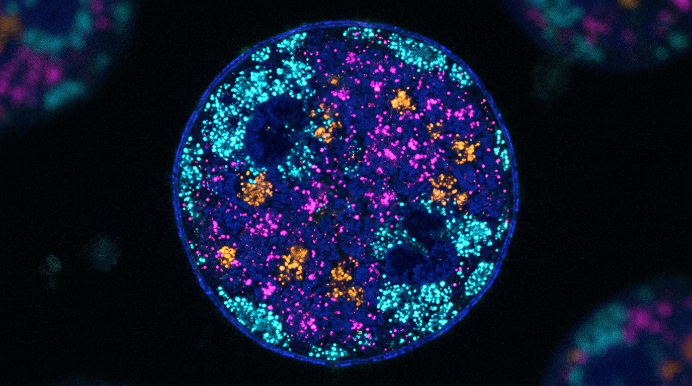
A newly developed spatial profiling methodology named epigenetic proximity hybridization reaction, or Epi-PHR, allows researchers to map combinations of histone modifications at targeted genomic loci in single cells while preserving three-dimensional chromatin architecture. Reporting in Nature Methods, an investigative team led by Yanbo Chen, Jonathan S. D. Radda, Myles H. Alderman III, Shengyan Jin, Yubao Cheng, Yuan Zhang, Miao Liu, Andrew Z. Xiao, and Siyuan Wang addressed a critical challenge in single-cell genomics: existing sequencing and spatial omics technologies have struggled to resolve locus-specific combinations of epigenetic marks without disrupting the native spatial folding of chromatin. By coupling locus-targeted fluorescence in situ hybridization with an engineered proximity-guided DNA assembly cascade, the nondisruptive imaging platform detects specific chromatin marks in situ across hundreds of genomic targets within individual nuclei. Crucially, the technology preserves native nuclear structures, enabling investigators to track how distinct chemical modifications interface with three-dimensional chromatin folding at single-gene and single-chromatin-copy resolution.
The molecular mechanism of Epi-PHR operates through a tightly regulated proximity reaction that converts spatial closeness between an epigenetic mark and a specific genomic locus into a fluorescent readout. In fixed and permeabilized cells, primary antibodies selectively bind to the epigenetic modification of interest, followed by incubation with biotinylated secondary antibodies. Samples are post-fixed and embedded within a protective polyacrylamide hydrogel matrix before being incubated with locus-specific fluorescence in situ hybridization probes. The proximity assay is driven by two DNA hairpin oligonucleotides: a PH1 hairpin attached to genomic probes via a docking adaptor, and a biotinylated PH2 hairpin linked to secondary antibodies through streptavidin. Adding an activator oligonucleotide opens the hairpin stem of PH1, which subsequently unzips the adjacent PH2 hairpin. A fluorescent dye-labeled probe designated H1 then hybridizes to the opened end of PH2, followed by sequential incubation with a second dye-labeled probe, H2. This controlled two-step hybridization delivers an exact twofold signal amplification rather than uncontrolled chain growth, while a distinct fluorophore on the genomic probe independently visualizes the three-dimensional locus coordinates.
A persistent technical hurdle during initial assay development was background signal caused by elevated activator concentrations stochastically opening PH2 hairpins away from target loci. The researchers resolved this issue by coupling hydrogel embedding with post-reaction enzymatic clearing using proteinase K. Following fluorescent signal generation, samples were treated with proteinase K to digest antibodies and release uncoupled PH2 hairpins, retaining only assemblies physically anchored to genomic FISH probes via PH1. To ensure unreacted PH1 molecules did not capture liberated PH2 hairpins during digestion, a blocking oligonucleotide was introduced beforehand to cap unoccupied PH1 ends. Proof-of-concept tests targeting a 100-kilobase heterochromatic region on chromosome 1 in IMR90 human fibroblasts confirmed that proteinase K clearing sharply reduced whole-nucleus background, greatly increasing the signal-to-background ratio over standard immunofluorescence. In systematic negative controls omitting key components—including activator, H1, PH1, PH2, secondary antibody, or streptavidin—foci signals were completely abolished, verifying that fluorescent readout depends strictly on complete, proximity-guided assembly.
To evaluate genomic sensitivity and spatial resolution boundaries, the team scaled FISH probe libraries across progressively smaller regions of chromosome 1 in diploid IMR90 cells. Probe sets ranged from 100 kilobases containing 2,219 probes down through 50, 20, 10, 5, and 2 kilobases using 52 probes, maintaining linear scaling between probe count and trimethylated histone H3 Lys9 (H3K9me3) peak coverage from published chromatin immunoprecipitation sequencing data. Distinct Epi-PHR foci colocalized with genomic FISH spots across all tested resolutions, yielding approximately two FISH foci per diploid cell. Although the signal-to-background ratio gradually declined as target spans narrowed, detection efficiency remained notably high, recording 99 percent at 100 kilobases and 67 percent at 2 kilobases. Furthermore, 81 percent of cells displayed at least one detectable Epi-PHR spot at 2-kilobase resolution. This sensitivity markedly exceeded existing benchmarks for spatial epigenomic methods, including epigenomic MERFISH, which achieves roughly 35 percent detection at 4-kilobase resolution, and single-cell CUT&Tag, which captures approximately 8 percent of targets at comparable genomic scales.
The analytical specificity of the system was rigorously challenged by targeting active epigenetic marks within the same 2-kilobase heterochromatic region on chromosome 1. Probing for acetylated histone H3 Lys27 (H3K27ac), phosphorylated RNA polymerase II Ser2 (Pol II S2P), or non-specific rabbit IgG controls produced minimal signal compared to H3K9me3. Low levels of residual active marks were suspected to arise from trans-interactions occurring when distant euchromatic regions transiently contacted the probed locus in three-dimensional space. Treating cells with the transcription elongation inhibitor flavopiridol to deplete Pol II S2P or with A485 to inhibit p300/CBP histone acetyltransferases further suppressed these residual signals, indicating that background was driven partly by physical trans-contacts. In addition, targeting an alternative 100-kilobase region with lower endogenous H3K9me3 density produced significantly reduced Epi-PHR signals. Based on probe, linker, and activator dimensions totaling 180 nucleotides, the researchers calculated an upper physical proximity threshold of 60 nanometers, emphasizing that Epi-PHR inherently detects three-dimensional spatial proximity rather than strictly linear cis-arrangements.
To achieve high-throughput multi-loci detection, Epi-PHR was integrated with DNA multiplexed error-robust fluorescence in situ hybridization (DNA MERFISH), enabling single-cell profiling of 200 gene loci simultaneously. Primary probes designed against these 200 genes contained a common PH1 docking sequence alongside locus-specific barcodes assigned a 36-bit Hamming-weight-2 code. Through 18 rounds of two-color imaging, sequential hybridization and chemical cleavage of fluorescent readout probes decoded locus coordinates across individual nuclei. In cultured IMR90 cells, Epi-PHR measurements of active H3K36me3 and repressive H3K27me3 demonstrated strong reproducibility between biological replicates and clear divergence from non-specific IgG controls. Signal rates correlated strongly with bulk ChIP-seq profiles, yielding Pearson correlation coefficients of 0.84 for H3K36me3 and 0.62 for H3K27me3, while capturing roughly 27 percent and 18 percent of bulk ChIP-seq peaks per cell, respectively. In parallel tests, epigenomic MERFISH produced a much lower ChIP-seq correlation for H3K27me3 (r = 0.15) and detected only 5 percent of bulk repressive peaks per cell, underscoring Epi-PHR’s superior capacity to profile compacted heterochromatin inaccessible to tagmentation enzymes.
The methodology was expanded to simultaneous dual-mark epigenetic profiling by engineering an orthogonal second set of hairpin oligonucleotides operating in parallel optical channels. Computational modeling guided the design of hairpin sequences that maintained comparable reaction kinetics while avoiding molecular cross-talk. By pairing secondary antibodies raised in different host species or conjugated directly to docking oligonucleotides, the researchers simultaneously mapped H3K36me3 and H3K27me3 across 200 genes in single cells. Single-mark enrichment predominated across most gene bodies, consistent with biological mutual exclusivity, whereas dual-enrichment was confined largely to topological domain boundaries. The researchers also validated dual-mark profiling in mouse brain hippocampus tissue sections, targeting H3K27ac and RNA polymerase II Ser5 phosphorylation across 200 genes. Anatomical segmentation of DAPI-stained images clearly separated cornu ammonis and dentate gyrus regions, demonstrating active mark enrichment at known cell-type marker genes derived from single-cell RNA-sequencing data, including Glis3 and Fam163b in the dentate gyrus and Lingo1 and Man1a in the cornu ammonis.
A major advance of Epi-PHR is its direct compatibility with chromatin tracing on identical single-cell targets. Targeting a 500-kilobase region on human chromosome 20 enriched for H3K27ac, the authors subdivided the sequence into 30 consecutive tracing segments of approximately 16.6 kilobases each. Probes carried both a shared PH1 docking sequence for Epi-PHR and locus-specific barcodes for tracing. Control experiments confirmed that neither polyacrylamide embedding nor proteinase K digestion altered native chromatin architecture: domain boundaries, loop anchors, and inter-loci spatial distances remained preserved and correlated strongly with Hi-C contact frequencies. Conversely, attempting to combine chromatin tracing with epigenomic MERFISH resulted in severe structural disruption. Because epigenomic MERFISH relies on Tn5 transposase cleavage that fragments double-stranded genomic DNA, subsequent denaturation and tracing steps caused tracing signals to disperse irregularly across nuclei, completely erasing folding patterns. This stark comparison highlights Epi-PHR’s non-cleaving mechanism, preserving genomic continuity for integrated structural and epigenetic analysis.
The combined imaging workflow was applied to examine allele-specific chromatin folding and epigenetic marking at the imprinted Meg3 locus within the Dlk1-Meg3 region in hybrid mouse trophoblast stem cells. Using cells derived from CAST and C57BL/6J parents, the authors coupled H3K9me3 Epi-PHR at Meg3 with chromatin tracing across a 1.2-megabase domain divided into thirty-four 35-kilobase bins. Alleles were distinguished using single-nucleotide polymorphism FISH probes targeting parental variant clusters flanking the locus. In agreement with the paternal imprinting status confirmed by allelic CUT&RUN, paternal Meg3 alleles exhibited significantly higher H3K9me3 signals and preferentially localized toward the repressive nuclear periphery. Chromatin tracing concurrently uncovered a maternal-specific domain boundary at the 5-prime edge of Meg3, matching maternal-specific CTCF insulator binding at the Meg3 differentially methylated region, along with elevated maternal isolation from neighboring chromatin.
Single-chromatin-copy measurements revealed an unexpected biological relationship between H3K9me3 enrichment and domain boundary formation. When traces were stratified into quintiles based on H3K9me3 signal, maternal Meg3 copies with higher H3K9me3 levels formed increasingly prominent domain boundaries rather than adopting an uninsulated paternal-like architecture, contradicting bulk sequencing models where H3K9me3 acts as a surrogate for boundary-disrupting DNA methylation. Conversely, paternal traces lacked domain boundaries across all quintiles, with lower H3K9me3 levels correlating with generalized domain decompaction. The authors note technical limitations, including current restrictions to dual-mark detection stemming from antibody species requirements, orthogonal hairpin engineering limits, and imaging color channels, alongside the need to interpret signals in light of 60-nanometer spatial trans-proximity. Nevertheless, by demonstrating high-resolution, non-destructive epigenetic profiling in single cells, Epi-PHR provides a powerful framework for deciphering how three-dimensional chromatin architecture coordinates with combinatorial histone modifications.
Subject of Research: Single-cell spatial profiling of combinatorial epigenetic marks and three-dimensional genome architecture
Article Title: Nondisruptive 3D profiling of combinations of epigenetic marks in single cells
Article References: Chen, Y., Radda, J. S. D., Alderman, M. H., III, Jin, S., Cheng, Y., Zhang, Y., Liu, M., Xiao, A. Z., & Wang, S. (2026). Nondisruptive 3D profiling of combinations of epigenetic marks in single cells. Nature Methods, 23(11), 2155-2168. https://doi.org/10.1038/s41592-026-03253-0
Image Credits: AI Generated
DOI: 10.1038/s41592-026-03253-0
Keywords: epigenetics, spatial biology, single-cell imaging, chromatin architecture, histone modifications, genomics, MERFISH, chromatin tracing, Nondisruptive, profiling, combinations, epigenetic
News Source: Juliet Wilcox. (November 6, 2026). Nondisruptive Spatial Imaging Maps Combinations of Epigenetic Marks in Single Cells. Scienmag.