Electroacupuncture applied at the Zusanli acupoint significantly mitigates joint inflammation and protects articular cartilage in a mouse model of knee osteoarthritis through a defined central-to-peripheral neural circuit driven by cholinergic neurons in the dorsal motor nucleus of the vagus, according to a laboratory study published in Anesthesiology and Perioperative Science. The investigation, conducted in eight-week-old male mice, demonstrated that electrical stimulation at the ST36 acupoint selectively activates choline acetyltransferase-positive cholinergic neurons situated within the dorsal motor nucleus of the vagus in the brainstem. By combining in vivo calcium fiber photometry, chemogenetic excitation and inhibition, subdiaphragmatic vagotomy, transcriptomic sequencing, and histopathology, the team mapped the neuroanatomical axis bridging brainstem activation to joint-level anti-inflammatory control in experimental osteoarthritis.
Knee osteoarthritis represents the most prevalent degenerative joint disorder and stands as a leading contributor to chronic physical disability and diminished quality of life among elderly populations worldwide. Epidemiological assessments cited by the researchers indicate that global osteoarthritis cases reached approximately 595 million individuals by 2020, with prevalence rates expected to sustain an upward trajectory due to demographic aging, physical trauma, metabolic syndromes, and obesity. Despite this substantial clinical burden, conventional management strategies exhibit significant therapeutic constraints: long-term pharmacological interventions frequently provoke systemic adverse effects, surgical replacements involve high operative risks and procedural complexity, and physical modalities often demonstrate limited clinical effectiveness. In response to these treatment gaps, traditional Chinese medicine acupuncture, particularly modern electroacupuncture utilizing controlled alternating electrical stimulation, has garnered increasing scientific interest. Electroacupuncture delivers electrical currents to deep nerves, muscles, and fascia with precise parameter controllability, providing sustained therapeutic outcomes across diverse indications.
To investigate the underlying central nervous system mechanisms, the authors established a standardized model of knee osteoarthritis using eight-week-old male C57BL/6J mice, alongside transgenic FosCreER (TRAP2) and TRAP2-Ai47 mouse strains housed under controlled environmental conditions. Unilateral osteoarthritis was induced through an intra-articular injection of monosodium iodoacetate into the right knee joint space at a dosage of one milligram in fifty microliters of sterile saline, followed by manual flexion and extension to ensure dispersion, while control animals received sterile saline. After two weeks of model stabilization, experimental mice were assigned to sham acupuncture or active electroacupuncture administered under isoflurane anesthesia. For the active electroacupuncture group, stainless-steel positive needles were inserted four millimeters deep into the right Zusanli acupoint—located four millimeters below the knee joint and five millimeters lateral to the anterior tibial tubercle—with negative needles positioned one millimeter aside. Electrical stimulation was delivered daily for fourteen consecutive days using an alternating frequency of 2/100 Hertz at two milliamperes for thirty minutes per session.
Comprehensive histopathological evaluations of knee joint specimens following the fourteen-day intervention revealed that electroacupuncture treatment substantially reversed monosodium iodoacetate-induced joint degeneration, whereas non-electrical manual acupuncture produced negligible therapeutic benefits. Histological staining with hematoxylin and eosin, alongside Safranin O/Fast Green and collagen type II immunohistochemistry, demonstrated marked restoration of articular cartilage surface smoothness, significant replenishment of depleted glycosaminoglycan content, and increased proportions of collagen type II-positive chondrocytes within the articular matrix. Quantitative assessments confirmed profound reductions in Osteoarthritis Research Society International histopathology scores and cartilage lesion thickness in electroacupuncture-treated rodents compared to untreated arthritic cohorts. Furthermore, enzyme-linked immunosorbent assays performed on synovial tissue extracts revealed that electroacupuncture suppressed pathologically elevated levels of primary pro-inflammatory cytokines, specifically interleukin-1 beta, interleukin-6, and tumor necrosis factor-alpha, restoring them near baseline levels observed in healthy control animals.
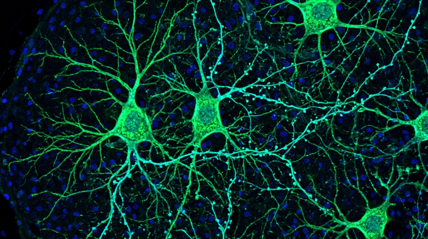
To identify which central nervous system structures mediate these peripheral anti-inflammatory effects, the researchers conducted whole-brain immunofluorescence screening targeting the immediate early gene c-Fos, a widely established cellular marker of neuronal activation. Following electroacupuncture stimulation at the ST36 acupoint, elevated c-Fos expression was detected across multiple cerebral regions, with the dorsal motor nucleus of the vagus displaying the most robust activation response. Multiplex immunofluorescence co-staining of dorsal motor nucleus sections for c-Fos alongside neurochemical markers—including choline acetyltransferase, tyrosine hydroxylase, calcium/calmodulin-dependent protein kinase II alpha, and glutamate decarboxylase 1—revealed that activated neurons were predominantly positive for choline acetyltransferase. To verify dynamic activation in real time, the investigators stereotaxically delivered an adeno-associated viral vector encoding the calcium indicator GCaMP6s under a choline acetyltransferase promoter into the left dorsal motor nucleus. In vivo fiber photometry recordings during electroacupuncture revealed significant increases in intracellular calcium fluorescence transients and response amplitudes.
Having identified this responsive cholinergic population, the scientists examined whether selective activation of dorsal motor nucleus neurons could reproduce the therapeutic efficacy of electroacupuncture in arthritic mice without peripheral needle stimulation. Using FosCreER (TRAP2) mice, the team stereotaxically injected adeno-associated viral vectors encoding double-floxed inverted orientation Gq-coupled designer receptors tagged with mCherry into the bilateral dorsal motor nucleus relative to Bregma coordinates. Chemogenetic activation via systemic clozapine-N-oxide administration induced robust c-Fos expression within targeted brainstem neurons, reproducing the structural and biochemical benefits of physical electroacupuncture. Arthritic mice undergoing chemogenetic activation showed marked preservation of articular cartilage thickness, restored glycosaminoglycan content, normalized Osteoarthritis Research Society International scores, and increased collagen type II-positive chondrocytes. In addition, synovial enzyme-linked immunosorbent assays demonstrated significant suppression of interleukin-1 beta, interleukin-6, and tumor necrosis factor-alpha, accompanied by elevated synovial dopamine levels, whereas control vectors lacking the activating receptor yielded no protection.
To establish the necessity of cholinergic dorsal motor nucleus neurons in electroacupuncture-mediated joint protection, the researchers performed loss-of-function experiments using inhibitory chemogenetics in wild-type mice. Animals received bilateral stereotaxic microinjections of recombinant adeno-associated viruses expressing Gi-coupled designer receptors under the control of the choline acetyltransferase promoter. Prior to daily thirty-minute sessions of electroacupuncture at ST36, mice were administered intraperitoneal clozapine-N-oxide injections for one week. Immunofluorescence confirmed successful silencing through the marked diminution of c-Fos expression in cholinergic neurons. Functional inhibition of these neurons completely abolished the therapeutic benefits of electroacupuncture: cartilage lesion severity worsened, glycosaminoglycan depletion persisted, and Osteoarthritis Research Society International damage scores remained high. Crucially, synovial enzyme-linked immunosorbent assays demonstrated that chemogenetic silencing prevented electroacupuncture from lowering interleukin-1 beta, interleukin-6, and tumor necrosis factor-alpha levels, while abolishing the elevation of synovial dopamine, confirming that dorsal motor nucleus cholinergic activity is essential for electroacupuncture efficacy.
Because the dorsal motor nucleus serves as the central origin for efferents of the vagus nerve, the researchers investigated whether peripheral vagal innervation is required to convey anti-inflammatory signals from the brainstem to the arthritic joint. To address this connection, the team performed bilateral subdiaphragmatic vagotomies in adult mice, exposing the subdiaphragmatic esophagus below the xiphoid process, transecting the posterior vagal trunks under a surgical microscope, and resecting a two-to-three-millimeter segment to prevent regeneration. In vagotomized arthritic mice, daily electroacupuncture at ST36 completely failed to reduce synovial levels of interleukin-1 beta, interleukin-6, and tumor necrosis factor-alpha. Furthermore, subdiaphragmatic vagotomy eliminated the electroacupuncture-induced elevation of dopamine within synovial tissue. These surgical results demonstrated that intact subdiaphragmatic vagal pathways projecting toward peripheral targets, specifically the adrenal medulla, are indispensable for converting central cholinergic stimulation into peripheral dopamine secretion and joint-level anti-inflammatory actions in knee osteoarthritis.
To uncover the molecular signaling cascades modulated within joint tissue following electroacupuncture, the authors performed high-throughput Illumina RNA sequencing on synovial tissue from control, osteoarthritic, sham-treated, and electroacupuncture-treated mice. Bioinformatic analysis using Kyoto Encyclopedia of Genes and Genomes pathway enrichment revealed significant transcriptional reprogramming, highlighting marked downregulation of the phosphoinositide 3-kinase–Akt signaling pathway in electroacupuncture-treated synovium. Quantitative real-time polymerase chain reaction confirmed this molecular suppression, revealing significant reductions in pathway-related genes that were elevated in arthritic synovium, including insulin receptor substrate 1, phosphoenolpyruvate carboxykinase 1, and collagen type II alpha 1, alongside collagen types IV, VI, and IX alpha chains.
In their overall synthesis, the authors demonstrated that electroacupuncture at the Zusanli acupoint engages an integrated dorsal motor nucleus–vagus nerve–adrenal axis, elevating local dopamine to suppress joint inflammation and reverse cartilage degradation in experimental osteoarthritis. The authors contextualized these results within broader neuroimmunology, noting that while dopamine acts through dopamine receptors expressed on synovial fibroblasts to inhibit cytokine cascades, the specific receptor subtypes mediating these actions remain to be characterized. The authors also emphasized that these findings derive from chemically induced knee osteoarthritis in male mouse models, meaning that direct clinical translation requires large-scale multicenter human clinical trials to verify whether identical vagal-adrenal pathways operate in human patients. Delineating these central cholinergic brainstem circuits provides a foundational mechanistic framework for targeted neuromodulation therapies in joint disease.
Subject of Research: Central neural mechanisms of electroacupuncture in knee osteoarthritis
Article Title: Activation of DMV cholinergic neurons mediates the anti-inflammatory effects of electroacupuncture in knee osteoarthritis mice
Article References: Lu, S., Wang, B., Zhou, Y., Luo, H., Zhang, S., Zhan, Q., Jiao, Y., Yu, W., Xiao, J., Gao, P., & Yang, L. (2026). Activation of DMV cholinergic neurons mediates the anti-inflammatory effects of electroacupuncture in knee osteoarthritis mice. Anesthesiology and Perioperative Science, 4(1), Article 34. https://doi.org/10.1007/s44254-026-00186-x
Image Credits: AI Generated
DOI: 10.1007/s44254-026-00186-x
Keywords: electroacupuncture, knee osteoarthritis, dorsal motor nucleus of the vagus, vagus nerve, dopamine, neuroimmunology, anti-inflammatory, cartilage repair, Activation, cholinergic, neurons, mediates
News Source: Ophelia Keating. (November 6, 2026). Brainstem Cholinergic Neurons Drive Electroacupuncture Anti-Inflammatory Effects in Arthritic Mice. Scienmag.