


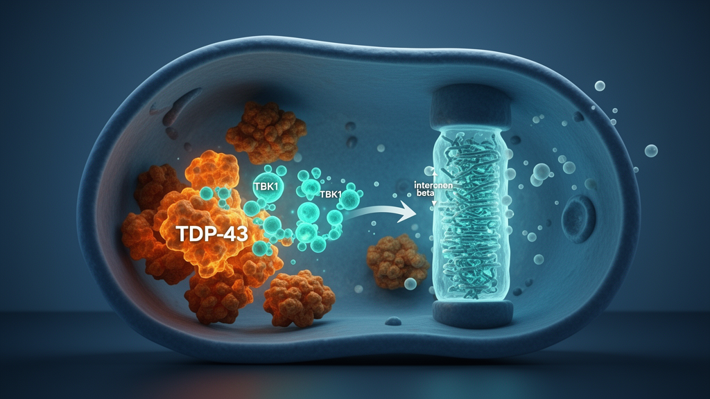
A kinase whose loss-of-function mutations cause familial amyotrophic lateral sclerosis and frontotemporal dementia has now been shown to clear the protein that defines those diseases through a route that bypasses autophagy entirely. In a study published in EMBO Reports, researchers led by Koji Yamanaka report that TANK-binding kinase 1, or TBK1, is switched on by the aggregation-prone monomeric form of TDP-43 and responds by inducing interferon-β, which in turn upregulates the immunoproteasome to degrade the offending protein in neurons, including human induced pluripotent stem cell-derived lower motor neurons. The work identifies an autophagy-independent proteostatic pathway that may explain why TBK1 mutations produce the cytoplasmic TDP-43 aggregates that are the pathological hallmark of ALS and FTD.
The team’s starting point was a paradox. TBK1 is already known to phosphorylate the autophagy receptors optineurin and p62, promoting clearance of damaged organelles and protein aggregates, and both OPTN and SQSTM1 are recognized ALS/FTD causative genes. Yet the authors hypothesized that if TBK1 is crucial for suppressing TDP-43 pathology, it should be activated in response to a monomeric TDP-43 burden. Using Neuro2a and NSC-34 cells transfected with either wild-type human TDP-43 or the 6M mutant, which carries six point mutations at the homo-dimerization and oligomerization interface and recapitulates key features of TDP-43 pathology, they found that phosphorylated TBK1, the active form of the kinase, was increased in cells exhibiting cytoplasmic TDP-43, particularly in cells expressing the 6M mutant, and that the phosphorylated kinase colocalized with cytoplasmic monomeric TDP-43.
Critical in vivo evidence came from postmortem human tissue. In spinal motor neurons from three sporadic ALS patients with TDP-43 pathology, the researchers observed colocalization of monomeric TDP-43 and phosphorylated TBK1, detected with a monomeric TDP-43-specific antibody. Colocalization was observed in 4 of 28, 3 of 13, and 5 of 26 motor neurons from the three ALS cases, while no motor neurons from three control subjects showed cytoplasmic colocalization. Overexpressed TBK1 was also shown to interact with monomeric TDP-43 by live-cell imaging and co-immunoprecipitation in a kinase activity-dependent manner, and DSP crosslinking followed by co-immunoprecipitation demonstrated that monomeric TDP-43 interacts with endogenous TBK1. Reducing endogenous TBK1 activity, either by siRNA-mediated knockdown or by treatment with the TBK1-specific inhibitor BX795, increased levels of monomeric TDP-43 and its phosphorylated form, indicating that the kinase normally restrains the accumulation of this pathological species.
The surprise came when the investigators tested whether the reduction in TDP-43 depended on autophagy. TBK1 overexpression did reduce TDP-43 levels in both the soluble and insoluble fractions of cell lysates, with a more pronounced effect on the 6M mutant than on wild-type TDP-43, and the effect required kinase activity, as ALS/FTD-linked kinase-dead TBK1 mutants such as R47H, G217R, R228H, R357Q, M559R, and Q565P failed to reduce TDP-436M levels. But when cells were treated with bafilomycin A1, a potent inhibitor of the autophagy-lysosome pathway, TDP-436M levels were still reduced by TBK1, even though the drug successfully blocked autophagic degradation, as shown by increased LC3-II levels. Proteasome inhibition by MG132, by contrast, blocked the reduction. These results pointed to a mechanism independent of the autophagy-lysosome pathway and dependent on the ubiquitin-proteasome system.
Because TBK1 is involved in cytokine secretion, the researchers next explored whether the kinase acts through humoral factors. Co-culture experiments revealed that neighboring cells overexpressing TBK1 reduced the protein levels of TDP-436M and phosphorylated TDP-43 in adjacent cells, and conditioned medium derived from TBK1-overexpressing cells reproduced the effect, ruling out physical cell-to-cell contact as the explanation. Knockdown of either Sting or Irf3 in the donor cells abolished the reduction of TDP-436M and p-TDP by TBK1-conditioned medium, and qPCR confirmed strong induction of Ifnb1 expression upon TBK1 overexpression, which was suppressed by Sting or Irf3 knockdown and was not induced by the kinase-dead mutants. Using two distinct siRNAs targeting Ifnb1, the team showed that knockdown of the cytokine in donor cells completely abolished the effect of TBK1-conditioned medium on TDP-436M and p-TDP levels, establishing IFNβ as the key cytokine responsible for TBK1-mediated clearance of the monomeric mutant. Inhibition of the JAK/STAT pathway, the canonical downstream signaling cascade of IFNβ, with the pan-JAK inhibitor tofacitinib also completely abolished the reduction, and knockdown of Ifnb1 exacerbated accumulation of TDP-43 and p-TDP, particularly in cells overexpressing TDP-436M.
To determine whether IFNβ alone is sufficient to reduce TDP-43 levels, the researchers tested recombinant IFNβ against a panel of TDP-43 mutants, including the monomeric mutants 6M and L27/28A, the nuclear localization signal-deficient ΔNLS, the aggregation-prone C173/175S, and the familial ALS-associated A315T and M337V. IFNβ treatment significantly reduced both monomeric mutants in the soluble and insoluble fractions, as well as p-TDP in the insoluble fraction, with a more pronounced effect on the monomeric mutants than on wild-type TDP-43 or ΔNLS. The effect was confirmed in stable cell lines, in the human neuronal cell line SH-SY5Y, and, importantly, in human iPSC-derived lower motor neurons generated from a healthy donor iPSC line using an NGN2-based differentiation protocol. The differentiated neurons expressed mature neuron markers such as MAP2 and neurofilament heavy chain and canonical lower motor neuron markers including HB9, ISL1, and choline acetyltransferase. IFNβ reduced monomeric TDP-43 in these post-mitotic neurons but not in undifferentiated iPSCs, and the effect was absent in HEK293 cells, suggesting that this regulatory mechanism may be specific to neurons.
The downstream mechanism involves the immunoproteasome, a specialized form of the proteasome complex that incorporates two immunoproteasome-specific regulatory subunits, PSME1 and PSME2, and three catalytic β-subunits, PSMB8, PSMB9, and PSMB10, facilitating the generation of peptides for MHC class I antigen presentation. Although IFNβ is not typically considered a canonical inducer of the immunoproteasome, previous studies have shown that type I interferons can induce immunoproteasome subunits in hepatocytes, epithelial cells, and fibroblasts. The researchers found that both IFNβ and TBK1-conditioned medium induced the expression of catalytic and regulatory immunoproteasome subunits in Neuro2a cells, and that IFNβ robustly induced PSMB8 and PSMB9 at the protein level in three neuronal cell lines, mouse primary spinal neurons, and iPSC-derived lower motor neurons, but not, or only faintly, in HEK293 cells, HeLa cells, and undifferentiated iPSCs. Using a PSMB8-specific fluorogenic substrate, they confirmed that relative immunoproteasome activity was increased by IFNβ or TBK1-conditioned medium and was not increased in the presence of the PSMB8-specific inhibitor ONX-0914. Treatment with ONX-0914 completely abolished the IFNβ-induced reduction of monomeric TDP-43, while the concentration used had minimal effects on the canonical proteasome, establishing the immunoproteasome as the degradation machinery responsible.
A second set of human tissue experiments revealed a potential vulnerability in the pathway. IFNβ binds to the interferon α/β receptor, a heterodimer composed of IFNAR1 and IFNAR2, and the researchers found that IFNAR1 is expressed in NeuN-positive neurons, including ChAT-positive spinal motor neurons and CTIP2-positive layer V cortical neurons, in wild-type mice. In the controls, strong IFNAR1 signals were observed in motor neurons, but IFNAR1 fluorescence intensity in motor neurons from ALS patients was significantly lower than in controls. Furthermore, comparing IFNAR1 intensity between TDP-43 pathology-negative and pathology-positive motor neurons within ALS patients, the researchers found that IFNAR1 expression was lower in pathology-positive neurons. The analysis used a linear mixed-effects model with diagnosis as a fixed effect and donor as a random effect, and the authors caution that the limited number of donors in the subgroup comparison means the results should be interpreted with care. The researchers also found that thapsigargin-induced ER stress reduced IFNAR1 levels in NSC-34 cells and iPSC-derived lower motor neurons, suggesting that ER stress, which is known to occur in ALS motor neurons, may contribute to the receptor’s downregulation and thereby impair protective type I interferon signaling during disease progression.
To test the pathway’s relevance in vivo, the team turned to SOD1G93A transgenic ALS model mice. They found that while canonical proteasome β-subunits Psmb6 and Psmb7 were downregulated in the lumbar spinal cord of the ALS mice at disease onset, 3.5 months of age, the immunoproteasome β-subunits Psmb8 and Psmb9 were upregulated. Laser microdissection of motor neurons from whole lumbar spinal cords confirmed that these molecular alterations occurred in motor neurons themselves, with reductions in canonical proteasome subunits and an increase in the immunoproteasome subunit Psmb8, suggesting that immunoproteasome induction in motor neurons may serve as a compensatory mechanism in response to increased proteostatic stress. Heterozygous Tbk1 deletion, confirmed by a 50 percent reduction of Tbk1 mRNA, led to decreased type I interferon responses, as assessed by reduced expression of Isg15 and Oas1a, and impaired immunoproteasome induction evaluated by the expression of Psmb8 and Psmb9. The authors note that homozygous deletion of Tbk1 in mice is embryonically lethal, which is why the heterozygous model was used.
The functional consequence of that impairment was measurable protein accumulation. SOD1G93A/Tbk1 heterozygous mice had higher levels of Triton-insoluble polyubiquitinated proteins in the lumbar spinal cord than SOD1G93A mice alone, while levels of the autophagy marker p62 did not differ between the two genotypes, suggesting a stage-dependent shift between autophagy and immunoproteasome pathways. The authors propose that the TBK1-IFNβ-immunoproteasome axis represents an inducible and protective mechanism that mitigates proteostatic stress in neurons, and that impairment of this pathway, whether through deregulated TBK1 or reduced IFNAR1 expression, may contribute to the development of TDP-43 pathology in ALS and FTD. They acknowledge limitations, noting that the expression levels of immunoproteasome subunits and IFNβ in patient tissues remain uncharacterized, and that whether the immunoproteasome is protective throughout the overall ALS/FTD disease course remains an open question. Previous mouse studies have shown that single genetic manipulation of Tbk1 is insufficient to recapitulate TDP-43 pathology, and the authors suggest that TBK1 appears to affect ALS and TDP-43 pathology as a so-called second hit, playing important roles particularly under disease conditions. Further studies with immunoproteasome-deficient ALS/FTD models using animals and iPSC-derived neurons or organoids will be required to address the remaining questions.
Subject of Research: A TBK1-IFNβ-immunoproteasome pathway that clears aggregation-prone monomeric TDP-43 in ALS and FTD models
Article Title: TBK1 eliminates aggregation-prone monomeric TDP-43 through an IFNβ-immunoproteasome pathway in ALS models
Article References: Sakai, S., Oiwa, K., Iguchi, Y., Akcay, I. M., Watanabe, S., Komine, O., Nagata, N., Horiuchi, M., Sreedharan, J., Katsuno, M., & Yamanaka, K. (2026). TBK1 eliminates aggregation-prone monomeric TDP-43 through an IFNβ-immunoproteasome pathway in ALS models. EMBO Reports. https://doi.org/10.1038/s44319-026-00953-8
Image Credits: AI Generated
DOI: 10.1038/s44319-026-00953-8
Keywords: TBK1, TDP-43, ALS, frontotemporal dementia, interferon-beta, immunoproteasome, autophagy, iPSC-derived motor neurons, SOD1G93A mice, IFNAR1, proteostasis, ER stress
News Source: Drew Townsend. (October 30, 2026). TBK1 clears aggregation-prone TDP-43 via an IFNβ-immunoproteasome pathway in ALS models. Scienmag.



