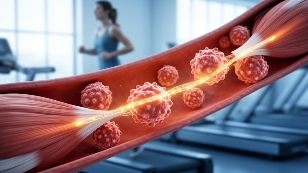
Exercise leaves a measurable imprint on the immune system, and a new study in Cardiovascular Diabetology traces that imprint down to individual genes and cell states in type 2 diabetes. The research, published on 24 October 2026, combines population health data, human single-cell sequencing and mouse experiments to ask how exercise-responsive genes connect T-cell metabolism with cardiovascular and cardiac adaptation in diabetes.
The team first turned to the National Health and Nutrition Examination Survey (NHANES), a large United States health survey. Higher physical activity was associated with lower prevalence of type 2 diabetes and more favorable glycemic profiles. Among participants who already had diabetes, higher physical activity was also associated with lower cardiovascular mortality. The authors stress that these are observational associations from survey data, not proof that exercise causes these outcomes, but they motivated the deeper molecular analysis that followed.
To identify which genes respond to exercise, the researchers performed an exploratory cross-species analysis of human and mouse skeletal-muscle transcriptomes. This yielded 18 exercise-responsive candidate genes. Among them, one stood out: JUNB, a transcriptional regulator whose expression varied along inferred T-cell pseudotime, a computational trajectory that orders single cells along a differentiation path. The team proposes JUNB as a candidate for further mechanistic investigation rather than a validated therapeutic target.
The human single-cell component used a peripheral blood mononuclear cell dataset. Compared with healthy controls, T cells from patients with periodontitis and diabetes mellitus showed lower TrainingScores, a metric derived from exercise-response signatures, and higher transcriptome-based scores for phenylalanine metabolism. The analysis also detected altered inferred communication between the molecules THBS1 and CD47 in these cells. These findings come from a disease group with both periodontal and metabolic inflammation, so they describe an association in a specific patient population rather than a general effect of exercise in diabetes.
The animal work focused on db/db mice, a widely used model of type 2 diabetes. After exercise training, splenic single-cell RNA sequencing identified eight distinct T-cell states. A pooled-cell analysis found a higher mean exercise-response score in regulatory T cells, known as Tregs, after exercise. Flow cytometry confirmed shifts in cell composition: the trained mice had higher proportions of Foxp3-positive CD4-positive regulatory T cells and lower fractions of pro-inflammatory CD4-positive T cells.
Cardiac responses were assessed with two independent mouse heart RNA-seq datasets. The comparison revealed partially concordant pathway-level responses across the two datasets, meaning some exercise-linked transcriptional pathways moved in the same direction in both, while others did not. The authors also note changes in cardiac morphological features in the exercise-trained db/db mice, alongside the transcriptional changes.
The overall conclusion is carefully bounded. Exercise-responsive candidate genes showed tissue- and cell-state-dependent expression patterns, and exercise-trained diabetic mice displayed changes in T-cell proportions together with cardiac transcriptional and morphological features. The study does not claim that exercise reverses diabetic heart disease in humans, nor that JUNB drives the observed effects. Instead, it maps where exercise-responsive signals appear across blood, spleen and heart tissue, and nominates specific molecular players for follow-up work.
Several limitations frame the findings. The human associations come from cross-sectional survey data, the single-cell human dataset involves patients with periodontitis and diabetes rather than a general diabetic cohort, and the mouse results derive from a single diabetic model with partially concordant cardiac datasets. The article was released early as a peer-reviewed, accepted version of record that remains subject to further edits. Within those limits, the work offers a concrete starting point for studying how exercise signals reach immune cells and the diabetic heart, with JUNB and the THBS1-CD47 signaling axis as the most clearly flagged leads.
Subject of Research: Exercise-responsive gene signatures connecting T-cell immunometabolism with cardiac adaptation in type 2 diabetes
Article Title: Exercise-responsive transcriptional signatures link T-cell immunometabolic remodeling with cardiovascular and cardiac adaptation in type 2 diabetes
Article References: Lin, W., Su, X., Wan, Z., Liao, Y., Huang, X., Lin, S., Yang, Y., Qiu, T., Fang, Z., Tian, J., & Zheng, Y. (2026). Exercise-responsive transcriptional signatures link T-cell immunometabolic remodeling with cardiovascular and cardiac adaptation in type 2 diabetes. Cardiovascular Diabetology. https://doi.org/10.1186/s12933-026-03420-9
Image Credits: AI Generated
DOI: 10.1186/s12933-026-03420-9
Keywords: type 2 diabetes, exercise, T cells, JUNB, single-cell RNA sequencing, regulatory T cells, NHANES, cardiac transcriptomics, db/db mice, immunometabolism, physical activity, cardiovascular mortality
News Source: Juliet Wilcox. (October 28, 2026). Exercise Reshapes T-Cell Gene Activity in Diabetes, Study Finds. Scienmag.