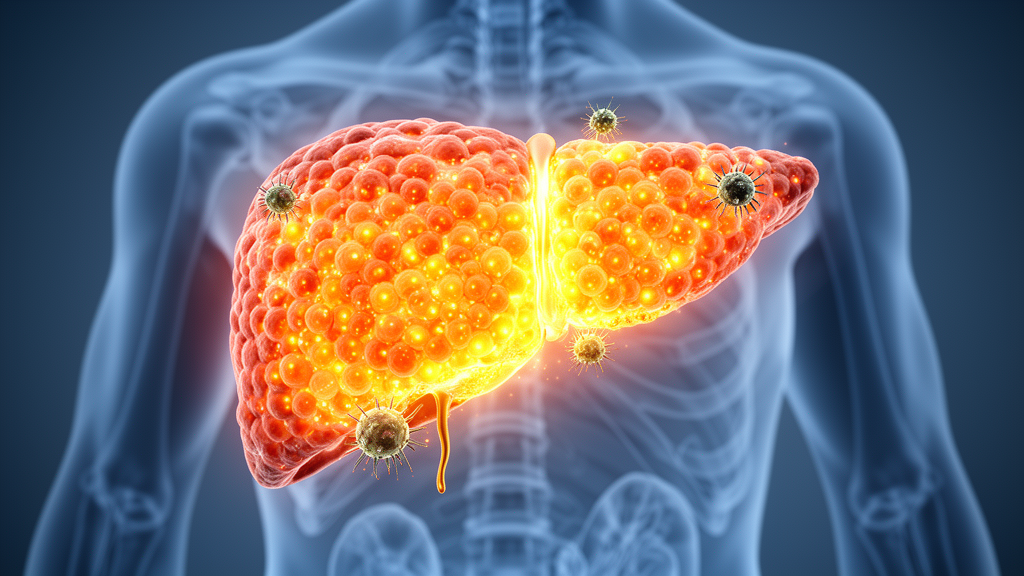
Hepatocellular carcinoma, the most common form of primary liver cancer, remains one of the deadliest malignancies in the world, ranking as the third leading cause of cancer-related mortality with a five-year survival rate that continues to disappoint clinicians and patients alike. Immune checkpoint inhibitors have transformed the treatment landscape for many cancers, and hepatocellular carcinoma is no exception, yet the sobering reality is that only roughly fifteen to twenty percent of patients achieve a durable response. Compounding the problem, physicians have lacked a reliable way to see, early and directly, whether the therapy is actually working inside a patient’s tumor. A new study published in Experimental & Molecular Medicine by Feng Xiong, Cheng Xu, Pei Wang, Ni Li and colleagues, led by corresponding authors Pengtao You, Dalong Ni and Bo Yu, now reports a strategy that tackles both problems at once, pairing a rational drug combination with a purpose-built positron emission tomography tracer that reads out the very molecular process the drugs are designed to trigger.
The conceptual breakthrough at the heart of the work lies in aligning the therapeutic pathway with the imaging target. Conventional response assessment during immunotherapy relies on CT or MRI, which measure lesion size and morphology, parameters that often change late and can even mislead clinicians during pseudoprogression. PET imaging offers a functional alternative, but existing tracers have their own shortcomings. The workhorse tracer fluorodeoxyglucose, known as FDG, suffers from variable uptake in hepatocellular carcinoma, while immunoPET tracers that visualize antigens such as GPC3, PSMA, HER2, PD-1 or PD-L1 confront a fundamental controversy: antigen expression is an unreliable predictor of which patients will respond. The researchers identified a deeper structural flaw in the current paradigm. Immunotherapy activates one target to kill cancer cells, while imaging visualizes a different target on the dying cells, creating a mismatch between what is treated and what is measured.
To close that gap, the team built the therapy and the imaging probe around a single molecule: SLC7A11, the functional subunit of the cystine-glutamate antiporter system xc-minus. This transporter imports extracellular cystine, the rate-limiting precursor for glutathione synthesis, and its suppression cripples the cellular antioxidant machinery. When SLC7A11 falls, cells become vulnerable to ferroptosis, an iron-dependent form of cell death driven by the accumulation of lipid peroxides. Crucially, both arms of the proposed combination therapy converge on this same molecule. Interferon-gamma released by activated CD8-positive T cells suppresses SLC7A11 and its partner SLC3A2, a mechanism established in earlier work showing that CD8-positive T cells regulate tumor ferroptosis during immunotherapy. Meanwhile, Olaparib, a clinically approved poly(ADP-ribose) polymerase inhibitor, has been shown to promote ferroptosis by repressing SLC7A11 even in cancers that are BRCA wild-type, converting single-strand breaks into lethal double-strand breaks and simultaneously sensitizing tumors to PD-L1 blockade by upregulating PD-L1 expression.
The therapeutic experiments were carried out in Hepa1-6 tumor-bearing C57BL/6J mice, which received intraperitoneal injections of an anti-PD-L1 antibody together with Olaparib every three days starting ten days after tumor inoculation. The results were striking. Tumor volume in the combination group fell to 155.4 plus or minus 69.5 cubic millimeters, compared with 361.6 plus or minus 153.3 cubic millimeters in the control group, a statistically significant reduction corresponding to roughly a 57 percent decrease. Immunohistochemical staining for Ki-67, a canonical marker of cell proliferation, dropped markedly in the treated tumors, while markers of DNA damage and oxidative injury, gamma-H2AX and 8-hydroxydeoxyguanosine, rose substantially. The combination did not merely add the effects of the two drugs; it synergized, and the pathological evidence pointed squarely at ferroptosis as the executing mechanism.
The mechanistic dissection reinforced that conclusion at every level examined. Transcriptomic analysis of Hepa1-6 cells revealed enrichment of immune system processes and oxidative stress genes, including Stat1, Irf1 and Ddit3, after interferon-gamma and combination treatment. Colony formation assays showed that Olaparib and interferon-gamma each inhibited clonogenic survival, with the combination producing synergistic killing. Reactive oxygen species, visualized by fluorescence microscopy with the probe H2DCFDA, climbed modestly with either agent alone but surged when the two were applied together. Western blotting confirmed significant downregulation of both SLC7A11 and GPX4, the glutathione-dependent selenoenzyme that normally detoxifies phospholipid hydroperoxides, in the combination group compared with either monotherapy. In the tumors themselves, glutathione was depleted most severely and malondialdehyde, a lipid peroxidation end product, reached its highest levels in the combination group, the biochemical signature of ferroptosis in full swing. Rescue experiments with the ferroptosis inhibitor Ferrostatin-1 reduced reactive oxygen species and reversed the therapeutic effect, sealing the mechanistic case.
The immune dependence of the regimen emerged as a second key finding. Immunohistochemistry revealed that both anti-PD-L1 and Olaparib monotherapies enhanced antitumor immune responses at day seven, with the combination producing the highest levels of interferon-gamma. By day fourteen, CD8-positive T cell infiltration had stabilized under anti-PD-L1 alone but continued to climb under Olaparib, reaching 11.13 plus or minus 2.28 percent positive area as a monotherapy and 13.02 plus or minus 2.19 percent in the combination, compared with 1.38 plus or minus 0.13 percent in controls. This suggests that the PARP inhibitor sustains T cell recruitment or activation in later stages of treatment, complementing the checkpoint blocker’s early boost. Most tellingly, when the same combination was tested in immunodeficient BALB/c nude mice, the therapeutic effect vanished entirely: tumors did not shrink, CD3 and CD8 staining was negligible across all groups, and neither tracer showed any differential uptake. The synergy, in other words, is fundamentally T cell dependent.
Onto this therapeutic platform the researchers grafted their imaging solution. The tracer, fluorine-18 labeled 5-fluoro-aminosuberic acid, abbreviated as F-18 FASu, targets system xc-minus and therefore reports on SLC7A11 expression. Unlike the older tracer F-18 FSPG, FASu shows greater specificity for system xc-minus and avoids interference from excitatory amino acid transporters, a property the team confirmed experimentally. The tracer was synthesized with a decay-corrected radiochemical yield of 18 plus or minus 6 percent, radiochemical purity above 99 percent and molar activity of 24 plus or minus 4 gigabecquerels per micromole. When mice were imaged on day eight of treatment, one hour after intravenous injection of 3.7 megabecquerels of the tracer, the combination group displayed a maximum standardized uptake value of 1.00 plus or minus 0.28, significantly lower than the control value of 1.76 plus or minus 0.48, and a tumor-to-background ratio of 14.31 plus or minus 2.87 versus 26.71 plus or minus 10.14. By contrast, FDG PET showed no statistically significant differences between groups, underscoring that the signal was specific to the ferroptosis pathway rather than a generic metabolic change.
The quantitative correlations lent further biological credibility to the imaging readout. Tumor SUVmax values correlated inversely with the pathological CD8-positive area, with a Pearson coefficient of minus 0.8095, and correlated positively with SLC7A11 expression measured by immunohistochemistry, with a coefficient of 0.8909. Cellular uptake assays mirrored the in vivo findings, with combination-treated cells accumulating less of the tracer. Taken together, these data establish F-18 FASu PET as a direct, non-invasive readout of the therapeutic mechanism itself: when the drugs work, SLC7A11 falls, ferroptosis advances, and the tracer signal dims accordingly. This is the essence of the theragnostic paradigm the authors propose, in which the same molecular target serves both the treatment and the assessment of that treatment, eliminating the mismatch that has plagued immunotherapy imaging.
The authors are careful to delineate the limitations that stand between this preclinical success and clinical practice. The study did not assess acute or chronic toxicity of the combination, and although both anti-PD-L1 antibodies and Olaparib are individually well characterized in the clinic, their combined toxicity profile requires dedicated investigation. The proposed T cell-interferon-gamma-SLC7A11-ferroptosis axis remains a working model consistent with the data but not yet validated by functional experiments such as T cell depletion or interferon-gamma neutralization. F-18 FASu itself is still at the preclinical stage, with toxicity, safety and human dosimetry studies yet to be conducted, and accurate response evaluation may require a baseline scan before therapy begins, adding clinical burden. Nevertheless, the convergence demonstrated here, in which a checkpoint inhibitor and a DNA repair inhibitor jointly drive cancer cells into ferroptosis while a targeted PET tracer watches the process unfold in living tissue, offers a genuinely new template for image-guided immunotherapy, one that could ultimately let oncologists know within days, rather than months, whether a liver tumor is surrendering to treatment.
Subject of Research: PET imaging of SLC7A11-mediated ferroptosis to monitor anti-PD-L1 and Olaparib combination therapy in hepatocellular carcinoma
Article Title: PET imaging enables direct evaluation of ferroptosis in HCC synergistically induced by anti-PD-L1 and Olaparib
Article References: Xiong, F., Xu, C., Wang, P., Li, N., Yang, Y., Zhang, B., Kwak, K., Song, S., You, P., Zhu, X., Ni, D., & Yu, B. (2026). PET imaging enables direct evaluation of ferroptosis in HCC synergistically induced by anti-PD-L1 and Olaparib. Experimental & Molecular Medicine. https://doi.org/10.1038/s12276-026-01860-7
Image Credits: AI Generated
DOI: 10.1038/s12276-026-01860-7
Keywords: hepatocellular carcinoma, ferroptosis, PET imaging, anti-PD-L1, Olaparib, SLC7A11, F-18 FASu, immunotherapy, PARP inhibitor, theranostics, molecular imaging, CD8 T cells
News Source: Nathaniel Bowman. (October 9, 2026). PET tracer lights up ferroptosis to track liver cancer immunotherapy in real time. Scienmag.