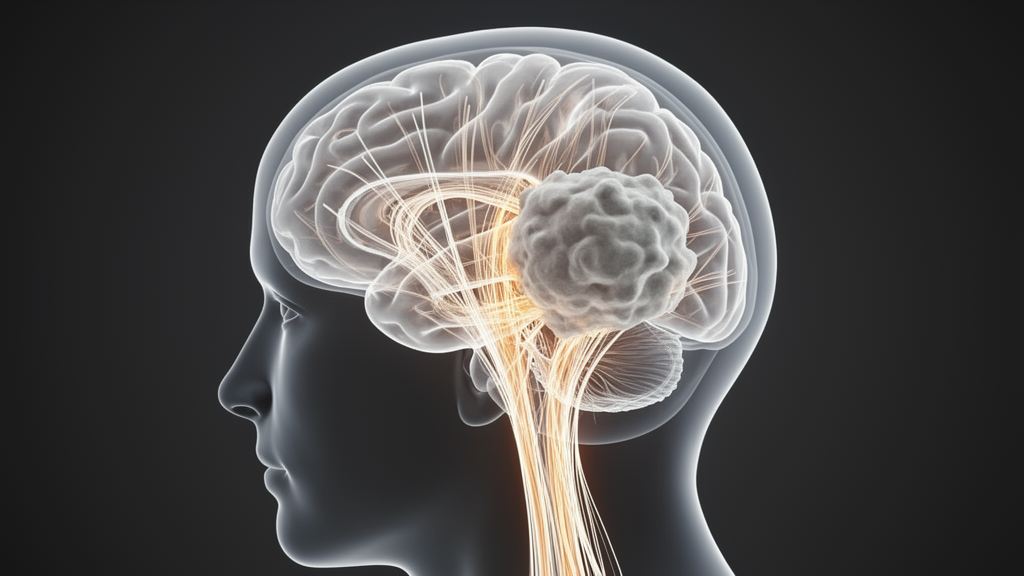
When neurosurgeons plan an operation for a brain tumor, they rely on a map of the patient’s motor pathways drawn from diffusion MRI. One of the most trusted anchors on that map is the corticospinal tract on the healthy side of the brain, the bundle of fibers that carries commands from the motor cortex down to the spinal cord. Because it sits far from the tumor, it has long been assumed to be a stable, patient-specific reference point against which the diseased hemisphere can be judged. A new longitudinal study challenges that assumption, showing that the healthy-side tract itself shifts position as a high-grade glioma progresses from first diagnosis to recurrence.
The study, published in the Journal of Neuro-Oncology by researchers at the Technical University of Munich, followed 29 adults with frontal-lobe grade 4 gliomas who underwent preoperative diffusion tensor imaging both before their initial surgery and again before surgery for documented recurrence. All patients had fully preserved motor strength, scoring 5 out of 5 on the Medical Research Council scale at both time points, which allowed the team to focus on changes in the brain’s wiring rather than on the consequences of weakness. The median interval between the two scans was just over ten months, a period during which most patients received radiotherapy, temozolomide chemotherapy, or both.
To reconstruct the corticospinal tract, the researchers used anatomically guided deterministic tractography, a technique that follows the preferred direction of water diffusion through white matter to trace fiber pathways. A manually drawn seed region at the level of the cerebral peduncle in the brainstem launched the tracking, the internal capsule served as an anatomical corridor constraint, and the precentral gyrus, the brain’s primary motor strip, defined the cortical endpoint. Crucially, the team ran the reconstruction twice, at fractional anisotropy thresholds of 0.10 and 0.15, to test whether their findings depended on how permissive the tracking algorithm was. Fractional anisotropy, or FA, is a measure of how directionally constrained water diffusion is; higher values generally indicate better-organized fiber bundles.
The first surprise was what did not change. When the researchers compared tract-averaged FA, tract length, and tract volume between the two time points, none of the six comparisons survived correction for multiple testing. A modest reduction in tract volume at the stricter threshold of 0.15 appeared in the raw statistics but dissolved once the false discovery rate correction was applied. In other words, by the standard quantitative yardsticks of tractography, the healthy-side motor pathway looked essentially the same at recurrence as it had at first presentation.
Position told a different story. By transforming each patient’s tract into a standardized brain template and computing the centroid, the geometric center of the tract, on every axial slice, the team could track where the bundle sat in three-dimensional space at each assessment. Across more than a hundred eligible slices, the tract showed a consistent anterior drift at recurrence, averaging 2.576 millimeters along the front-to-back axis at the permissive threshold and 2.078 millimeters at the stricter threshold. Dozens of individual slices remained statistically significant after rigorous correction, and the pattern held in sensitivity analyses using a nonparametric test. Side-to-side movement, by contrast, was small and inconsistent, averaging well under half a millimeter.
The researchers then compared their patients’ tracts against a fixed normative reference, the HCP842 atlas derived from 842 participants of the Human Connectome Project. Both the primary and recurrent tracts sat posterior to the atlas trajectory, but the recurrent tract was less posterior, sitting roughly two to three millimeters closer to the reference. This independently corroborates the within-patient finding: the healthy-side tract had genuinely moved forward over time, not merely drifted within the noise of the measurement. The authors are careful to note that these atlas comparisons are descriptive, since the atlas was generated with a different scanner protocol and tractography pipeline.
Why would the motor pathway on the opposite side of the brain shift at all? The study cannot answer that definitively, but the surrounding literature offers clues. Diffusion abnormalities have been documented in the apparently normal white matter contralateral to malignant gliomas, and serial imaging studies have shown progressive white matter changes during radiotherapy and chemotherapy. Whole-brain effects of tumor biology, treatment toxicity, edema dynamics, or compensatory plasticity could all contribute. Because surgery and adjuvant therapy occurred between the two scans in nearly all patients, the observed displacement cannot be attributed to recurrence alone; it reflects the cumulative journey of diagnosis, resection, and treatment.
The discrepancy between the spatial and the quantitative results is itself informative. The centroid analysis is exquisitely sensitive to distributed shifts in tract position, whereas the voxel-wise comparison, which tested every point in the standardized grid for reproducible gains or losses across patients, found nothing that survived correction. This suggests the displacement is spatially heterogeneous, varying in magnitude and direction from patient to patient, so that the group-level anterior shift emerges from averaging without any single voxel changing consistently. Individual trajectories indeed showed substantial variability superimposed on the group trend.
The clinical implications are immediate for anyone using the contralesional tract as a reference. If its position can move by two to three millimeters over the course of treatment, then importing a tract reconstruction from the initial surgery into a recurrent-case plan, or using the healthy side as a fixed baseline in longitudinal research, risks building the analysis on shifting ground. The authors recommend that longitudinal evaluations use contemporaneous tractography whenever it is available. At the same time, they caution that the functional meaning of the displacement remains unknown, since all patients had normal strength; future prospective studies with quantitative motor testing will be needed to determine whether the magnitude of the shift relates to fine motor performance, tumor proximity, or treatment exposure.
The study has clear limits. It was retrospective, single-center, and restricted to patients well enough to undergo paired imaging with successful tract reconstruction, which likely selected for a relatively favorable subgroup. Deterministic tractography remains sensitive to crossing fibers, seed placement, and threshold choices, and small spatial differences could partly reflect residual uncertainty in normalizing each brain to the template. Yet the consistency of the anterior shift across two tracking thresholds, two statistical approaches, and an independent normative reference makes the core message hard to dismiss: the brain’s geography is not static in glioma, and even the hemisphere that looks untouched is quietly rearranging itself as the disease and its treatment unfold.
Subject of Research: Longitudinal spatial displacement of the contralesional corticospinal tract in recurrent high-grade glioma measured with DTI tractography
Article Title: Longitudinal spatial changes of the contralesional corticospinal tract in high-grade glioma: a within-patient DTI tractography study
Article References: Longitudinal spatial changes of the contralesional corticospinal tract in high-grade glioma: a within-patient DTI tractography study. (n.d.). https://doi.org/10.1007/s11060-026-05823-w
Image Credits: AI Generated
DOI: 10.1007/s11060-026-05823-w
Keywords: glioma, corticospinal tract, diffusion tensor imaging, tractography, fractional anisotropy, brain tumor recurrence, neurosurgery, white matter, longitudinal study, MRI, neuro-oncology, motor pathways
News Source: Nathaniel Bowman. (October 9, 2026). The Brain’s Motor Highway Moves as Glioma Returns, Study Finds. Scienmag.