Sculpting of fish muscle requires genetic and mechanical information
Credit: Mechanobiology Institute, National University of Singapore
The diverse colours, shapes and patterns of fish are captivating. Despite such diversity, a general feature that we can observe in fish such as salmon or tuna once they are served in a dish like sushi, is the distinct ‘V’ patterns in their meat. While this appears to be genetically observed in the muscle arrangement of most fish species, how such a generic ‘V’ pattern arises is puzzling.
A team of researchers from the Mechanobiology Institute (MBI) at the National University of Singapore (NUS) investigated the science behind the formation of the ‘V’ patterns – also known as chevron patterns – in the swimming muscles of fish. The study focused on the myotome (a group of muscles served by a spinal nerve root) that makes up most of the fish body. These fish muscles power the fish’s side-to-side swimming motion and the chevron pattern is thought to increase swimming efficiency. The research team found that these patterns do not simply arise from genetic instruction or biochemical pathways but actually require physical forces to correctly develop. The findings of the study were published in the journal Proceedings of the National Academy of Sciences of the United States of America on 26 November 2019.
Friction and stress combine to shape patterns in fish muscle
The chevron pattern is not unique to salmon and tuna; it is also present in other fish species such as the zebrafish, as well as in some amphibian species like salamanders and frogs during development. The ‘V’ shape first appears in the somites – the precursor building blocks of the myotome, which forms the skeletal muscles. The somites typically form during the first few days of fish development or morphogenesis.
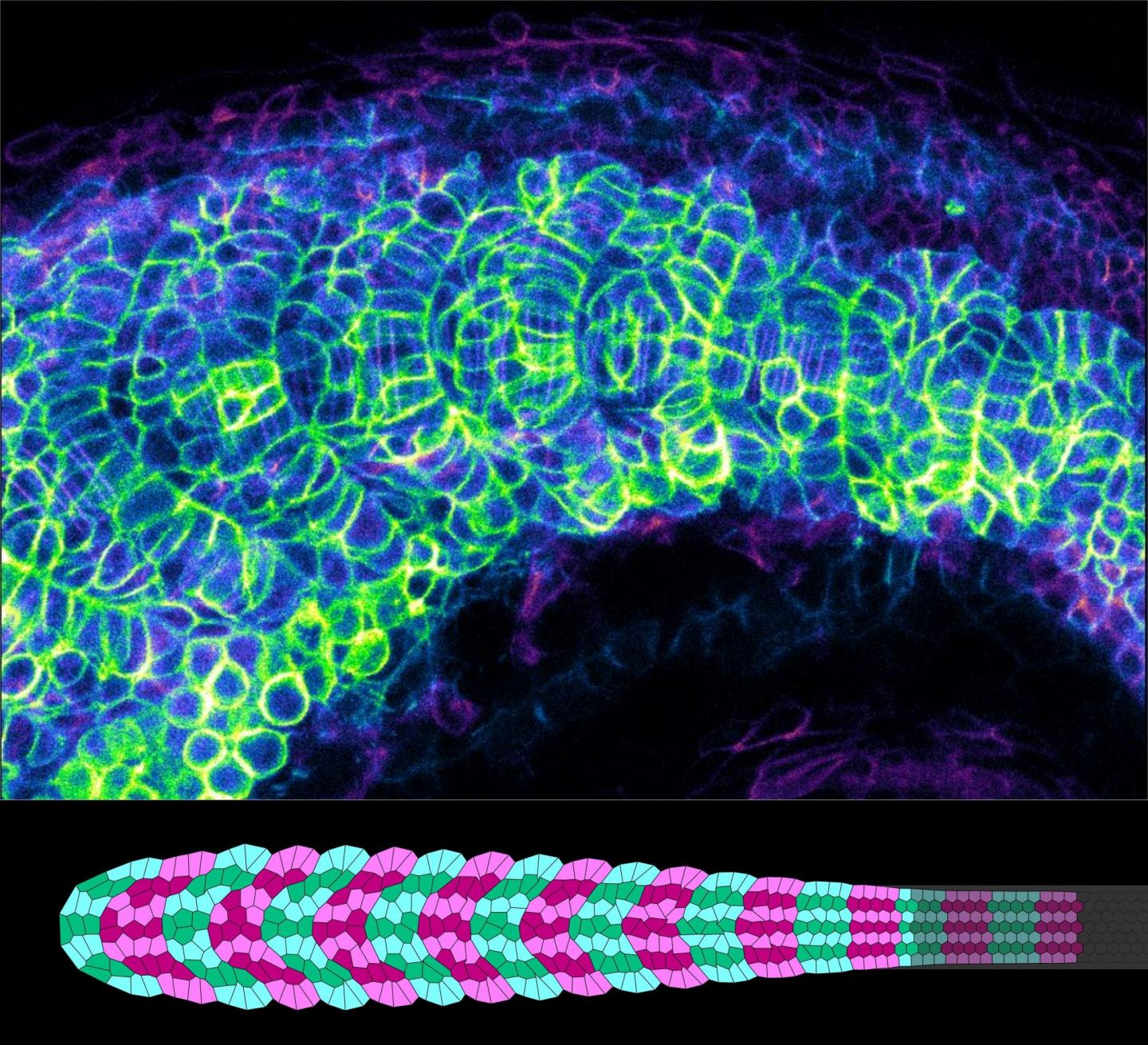
A team of scientists led by MBI Postdoctoral Fellow Dr Sham Tlili and Principal Investigator Assistant Professor Timothy Saunders studied chevron formation in the myotome of zebrafish embryos. Initially, each future developing myotome segment is cuboidal in shape. However, over the course of five hours, it deforms into a pointed ‘V’ shape. To find out how this deformation actually takes place, the team adopted a combination of different techniques – imaging of the developing zebrafish myotome at single cell resolution; quantitative analysis of the imaging data; and fitting the quantitative data into biophysical models.
Based on findings from their experimental as well as theoretical studies, the MBI scientists identified certain physical mechanisms that they thought might be guiding chevron formation during fish development.
Firstly, the developing myotomes are physically connected to other embryonic tissues such as the neural tube, notochord, skin and ventral tissues. The strength of their connection to these different tissues varies at different time points of myotome formation, and accordingly, different amounts of friction are generated across the tissue. Effectively, the side regions of the developing myotome are under greater friction than the central region. As new segments push the myotome forward, this leads to the formation of a shallow ‘U’ shape in the myotome tissue.
Secondly, cells within the future myotome begin to elongate as they form muscle fibres. The research team revealed that this transformation process generates an active, non-uniform force along certain directions within the somite tissue, which results in the ‘U’ shape sharpening into the characteristic ‘V’-shaped chevron. Lastly, orientated cell rearrangements within the future myotome help to stabilise the newly acquired chevron shape.
Deciphering the patterns guiding organ formation
Asst Prof Saunders, a theoretical physicist who applies physical principles to characterise biological processes that take place during development, said, “This work reveals how a carefully balanced interplay between cell morphology and mechanical interactions can drive the emergence of complex shapes during development. We are excited to see if the principles we have revealed are also acting in the shaping of other organs.”
It is common to attribute anything ‘appearance-related’ to the genetics of an organism. Through this study, the MBI scientists show how temporally and spatially varying biophysical forces play a role in determining the form of an organism.
###
Media Contact
Amal Naquiah
[email protected]
65-651-65125
Original Source
https:/
Related Journal Article
http://dx.
