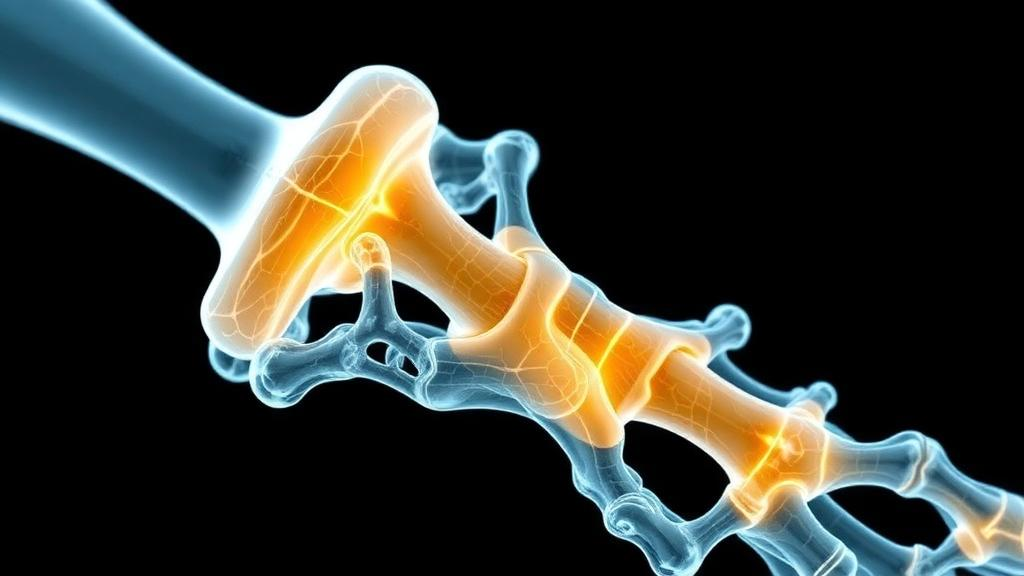
For decades, biomaterials scientists treated the immune response to an implanted bone scaffold as an enemy to be silenced. A new comprehensive review published in Materials Today Bio by Xiaonan Wang and Aobo Zhang argues that this long-standing assumption has been holding the field back, and that the future of bone regeneration lies not in avoiding inflammation but in choreographing it. Drawing together evidence from osteoimmunology, bioink chemistry and advanced fabrication, the authors lay out a framework in which three-dimensional bioprinting becomes the platform for actively steering immune cells through the precise sequence of states that natural bone healing requires.
The clinical problem is formidable. Bone defects caused by trauma, infection, tumour resection and congenital abnormalities are common, and while small injuries heal spontaneously, defects exceeding the local regenerative capacity, so-called critical-sized defects, often fail to close on their own. Autologous bone grafting remains the gold standard because it delivers osteogenic cells, inductive growth factors and a conductive matrix simultaneously, but it is limited by donor-site morbidity and restricted availability. Allografts are more available yet carry risks of immune rejection, infection and variable efficacy. What is needed, the review contends, are regenerative strategies that remain stable and reproducible inside the messy, inflamed microenvironment of a real defect.
Bioprinting offers an unusual degree of control over that environment. Unlike conventional fabrication, printing allows multiple materials, cells and bioactive signals to be arranged in three dimensions with defined pore architecture, compositional gradients and localised mechanical properties. Composite bioinks built from printable hydrogels such as gelatin methacryloyl (GelMA), alginate and hyaluronic acid methacrylate can be blended with inorganic phases including calcium phosphate, hydroxyapatite, beta-tricalcium phosphate and bioactive glass, balancing printability with an osteogenic milieu. Crucially, these same components release ions and present surface chemistry that shape the earliest host immune recognition of the implant, making the printer a tool for immunology as much as for structural engineering.
The biological rationale comes from the now well-established field of osteoimmunology. Bone repair unfolds in overlapping phases: an inflammatory phase dominated by neutrophils and pro-inflammatory M1-like macrophages that clear debris and pathogens; a proliferative phase in which macrophages shift toward reparative M2-like states, releasing factors such as TGF-beta and VEGF that recruit mesenchymal stem cells and drive angiogenesis; and a remodeling phase in which osteoblasts and osteoclasts, governed by the RANKL/OPG axis, convert woven bone into mechanically adapted lamellar bone. Simply suppressing the early inflammatory burst, the review stresses, can starve the repair process of the very signals that initiate cell recruitment and vascularisation. The goal instead is controlled, resolvable inflammation.
Material composition is one of the three main levers the authors identify. Strontium-substituted calcium silicate hydrate nanowires incorporated into a GelMA bioink, for example, released Sr, Ca and Si ions that nudged macrophages toward a repair-associated phenotype while supporting bone marrow stromal cell differentiation, producing substantial regeneration in rat critical-sized calvarial defects. Silicon-substituted calcium phosphate combined with methacrylated bone-derived extracellular matrix showed a striking temporal immune transition in vivo, from early pro-inflammatory activation to later anti-inflammatory resolution, linked to suppression of p38 MAPK signalling. Zinc-releasing ZIF-8 metal-organic framework particles delivered the anti-inflammatory polyphenol luteolin alongside Zn2+ ions, integrating immunomodulation, antioxidation and antibacterial action in a single printed construct.
Geometry itself can be immunologically active. Using digital light processing, researchers fabricated hydroxyapatite scaffolds identical in composition but differing only in pore size, and found that 600-micrometre pores best regulated macrophage behaviour through IFN-beta and HIF-1alpha-related signalling while improving vascularisation and bone formation. Even curvature matters: beta-tricalcium phosphate scaffolds engineered with negative Gaussian curvature suppressed macrophage Ras-MAPK/HIF-1alpha signalling and increased secretion of BMP-2 and VEGF, promoting vascularised bone ingrowth in a rabbit segmental defect model. Melt electrowriting adds another dimension, with ordered PCL microfibres and calcium phosphate coatings steering macrophages toward repair-associated states via PI3K/AKT and cAMP-PKA signalling, demonstrating that physical microarchitecture can substitute for exogenous drugs.
The most sophisticated strategies deliver bioactive cargo in stage-matched sequences. One printed system used coaxial bioprinting with sequential crosslinking to release interferon-gamma first, preserving the early inflammatory activation needed for repair initiation, before Laponite-derived magnesium and silicon ions gradually drove macrophages toward a reparative state, ultimately enhancing vascularised bone regeneration in calvarial defects. Interleukin-4 delivered from graphene oxide/black phosphorus nanointerfaces promoted CD206-positive macrophages while the materials themselves supported angiogenesis and osteogenesis. Exosome-based approaches add further nuance: macrophage-derived vesicles from mixed functional states outperformed those from purely M2-like macrophages in stimulating osteogenic signalling, underscoring the review’s warning that the simplified M1/M2 dichotomy cannot capture the continuum of immune states that real healing involves.
The authors are candid about translational hurdles. Every added component, whether living cells, recombinant cytokines, exosomes or decellularised matrix, increases batch variability, manufacturing complexity and regulatory burden. Sterilisation methods can silently alter hydrogel mechanics and even macrophage gene expression, while endotoxin contamination at low levels can confound the interpretation of a material’s intrinsic immunological effects. Degradation kinetics must be synchronised with tissue ingrowth: too fast and the scaffold loses support prematurely, too slow and the foreign-body response persists. The review advocates defining critical quality attributes that reflect immunomodulatory potency, not just scaffold geometry and compressive strength, and pursuing standardised platforms with patient-specific variation confined to a validated design space.
Looking forward, the field is moving toward spatiotemporal and even intelligent regulation. Microenvironment-responsive hydrogels that release therapeutic ions only under pathological acidity or oxidative stress, four-dimensional printing that changes scaffold architecture over time, and machine-learning frameworks that optimise bioink composition against immune as well as mechanical endpoints all feature in the review’s roadmap. Pathology-specific design receives particular emphasis: infected defects need preserved antimicrobial inflammation before resolution, diabetic defects require correction of hyperglycaemia-driven oxidative stress, and aged or osteoporotic bone demands restoration of senescence-impaired immune function rather than generic osteogenic stimulation. The overarching message is that the next generation of bone scaffolds should not simply contain more ingredients, but should speak the immune system’s own temporal language, guiding inflammation, vascularisation and remodeling in the order that nature intended.
Subject of Research: Immunoregulatory biomaterials and 3D bioprinting strategies for bone regeneration
Article Title: Advances in 3D bioprinting of immunoregulatory biomaterials for bone regeneration
Article References: Wang, X., & Zhang, A. (2026). Advances in 3D bioprinting of immunoregulatory biomaterials for bone regeneration. Materials Today Bio, 41, Article 103714. https://doi.org/10.1016/j.mtbio.2026.103714
Image Credits: AI Generated
DOI: 10.1016/j.mtbio.2026.103714
Keywords: 3D bioprinting, bone regeneration, osteoimmunology, macrophage polarization, bioinks, GelMA, biomaterials, critical-sized bone defects, exosomes, cytokine delivery, scaffold architecture, tissue engineering
News Source: Denise Maddox. (October 4, 2026). 3D Bioprinted Scaffolds That Coach the Immune System to Rebuild Bone. Scienmag.