


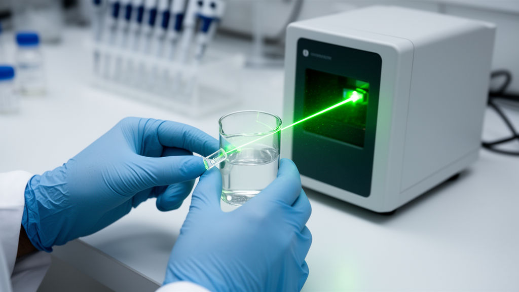
Protein self-assembly has just become a lot easier to watch as it happens. In an open-access protocol published in BMC Methods, researchers led by Christof M. Niemeyer at the Karlsruhe Institute of Technology describe a workflow that uses dynamic light scattering (DLS), a technique found in many biochemistry labs, to follow protein self-assembly in real time. Instead of measuring only the finished product, the method records the growth of assembling protein particles minute by minute, from the moment two complementary proteins are mixed until a stable nanoscale network has formed. The entire procedure, from sample preparation to data visualization and deposition, takes approximately eight hours and requires only small sample volumes and a standard benchtop DLS instrument.
The protocol addresses a genuine methodological gap. Endpoint characterization methods for protein assemblies are well established, but straightforward techniques to monitor protein self-assembly as it occurs have been lacking. According to the authors, this gap limits mechanistic understanding, hinders optimization of assembly conditions, and complicates quality control, particularly for systems that depend on precise stoichiometry or engineered interaction modules. Their answer is a continuous DLS workflow that records particle growth immediately after mixing and quantifies how the hydrodynamic diameter evolves over time, with a temporal resolution on the order of minutes.
DLS works by detecting time-dependent fluctuations in scattered laser light caused by the Brownian motion of particles in solution. The instrument converts the temporal autocorrelation function of these fluctuations into size information using established analytical models. The technique covers a broad size range, from 1 nanometer to 10 micrometers, and has long been applied to proteins, polymers, micelles, nanoparticles, and colloids. Although it is most commonly used to characterize samples at equilibrium, it can also capture dynamic processes: previous studies have used DLS to follow DNA origami assembly, crystal growth, protein aggregation, and fast kinetic processes with advanced modalities or stopped-flow coupling. What was missing, the authors argue, was a standardized and broadly accessible protocol for real-time monitoring of engineered protein self-assembly under controlled conditions.
The model systems at the heart of the protocol are all-enzyme hydrogels (AEHs), materials built from covalent SpyCatcher/SpyTag conjugation. Complementary enzymes equipped with these motifs crosslink spontaneously through isopeptide bond formation into dense yet hydrated three-dimensional networks that remain permeable for substrates and products. In solution, the initial crosslinking produces nanoscale hydrogel particles, which the authors call nanogels; upon concentration or drying, these coalesce into continuous macroscopic hydrogels. Such assemblies matter for industrial biocatalysis, where flow processes increasingly rely on gentle yet stable enzyme immobilization, and the balance of structural integrity and mass transport has already allowed AEHs to be implemented in durable microfluidic reactors, produced intracellularly, and advanced into monodisperse foam materials with excellent stability and flow performance.
The workflow itself has four main components. First, the individual proteins are prepared and quality-controlled: they must be at least 95 percent pure, free of high-molecular-weight contaminants, and freshly centrifuged or filtered to remove aggregates that would otherwise dominate the scattering signal, since intensity scales with the sixth power of particle diameter. Second, the self-assembly reaction is initiated by quickly mixing the proteins in a cuvette, and a sequence of repeated, identical DLS measurements begins immediately, with each measurement lasting 100 seconds and no waiting time between consecutive runs, continuing for at least two hours. Third, each individual protein is monitored alone over the same extended period, so that any size increase seen in the mixture can be distinguished from non-specific aggregation. Fourth, the resulting data are evaluated to extract growth profiles and assess assembly dynamics, with all measurements and metadata organized in a FAIR-compliant research data management workflow for transparent analysis and long-term reuse.
Data analysis proceeds along two routes. Monomodal, or cumulant, analysis fits a single exponential to the autocorrelation function and yields an intensity-weighted average hydrodynamic diameter, the Z-average, plus a polydispersity index (PDI) as an estimate of distribution width; it is preferred for monodisperse samples. Multimodal analysis fits multiple exponentials and generates an intensity-based distribution of particle sizes, which is better suited to the inherently polydisperse mixtures that arise during nanogel formation. The authors stress that Z-average values are intensity-weighted estimates, best suited for comparing relative growth trends rather than determining precise absolute particle sizes, particularly in polydisperse systems where datasets may yield high PDI values and resist standard cumulant analysis.
The applications demonstrate what the method can reveal. In an initial study, DLS quantified how protein concentration influences the dynamics of nanogel formation and how altering the stoichiometry of two SpyCatcher/SpyTag-interacting enzymes affects final particle size, enabling optimization of AEH synthesis conditions later implemented in continuous-flow biocatalytic reactors. A follow-up study compared engineered variants carrying different numbers of self-assembling motifs and determined optimal molar ratios for efficient network formation. More recently, the protocol was applied to proteins carrying non-covalent interaction modules: PDZ, SH3, and GBD domains. Qualitatively consistent with their reported binding affinities of roughly 8.0 micromolar for PDZ and 0.1 micromolar for SH3, the SH3-based system assembled more rapidly and reached a larger hydrodynamic diameter than the PDZ-based system, while both remained below the final sizes of the covalent SC/ST interaction. The GBD system, with an intermediate affinity of roughly 1 micromolar, displayed a much slower increase in diameter.
The numbers are striking. Linear regression of the early time points yielded initial particle growth rates of 0.12 nanometers per minute for the PDZ system, 1.97 nanometers per minute for SH3, and 0.06 nanometers per minute for GBD, at a total protein concentration of 0.5 millimolar and 25 degrees Celsius. The SH3 system reached an average particle size of 33 nanometers, PDZ approximately 22 nanometers, and GBD only 18 nanometers. Compared with covalent SC/ST assembly, which shows an initial growth rate of approximately 1.37 nanometers per minute and a particle size of about 50 nanometers, the non-covalent systems generally grew more slowly, with SH3 the exception. Interestingly, the order of growth rates did not strictly follow the expected trend based on binding affinities alone, suggesting that multivalent contacts inherent to the tetrameric enzyme architecture, steric constraints, and solvation effects also shape assembly kinetics and final nanogel size.
The protocol also exposes misbehaving components. In the GBD experiments, an unexpected increase in signal for one isolated protein, GDH-GBDL, suggests that this protein may undergo unspecific aggregation under the assay conditions, possibly due to insufficient stability of its tertiary structure during prolonged incubation, although the underlying cause cannot be conclusively assigned from DLS data alone. In the covalent PAD example, the individual PAD-SC protein showed behavior consistent with pronounced aggregation, with its PDI rising to the upper limit of 1, while the assembled mixture stabilized at a PDI of approximately 0.45, indicating that in this case self-assembly can reduce rather than amplify aggregation. For covalent systems, SDS-PAGE analysis of samples taken after the DLS measurements confirmed the formation of higher-molecular-weight conjugates corresponding to the expected assembly products.
The authors are candid about limitations. Very small oligomeric assemblies or weakly scattering systems may fall below the sensitivity range of standard benchtop instruments, and very fast assembly processes may exceed the effective temporal resolution imposed by manual mixing. Conditions approaching macroscopic gelation can impair data analysis through increased viscosity and reduced diffusion, and at high protein concentrations protein-protein interactions can invalidate the Stokes-Einstein relationship, meaning reported sizes reflect effective, interaction-influenced diffusion properties rather than true hydrodynamic radii of isolated species. The quantitative descriptors, including growth rates and apparent diameters, provide comparative characterization of assembly behavior rather than direct mechanistic insight, and additional orthogonal approaches would be needed to resolve underlying molecular mechanisms. Still, by pairing standardized time-resolved measurements with FAIR data management, the protocol offers an accessible, reproducible way to compare interaction modules, protein variants, and experimental conditions, and the authors expect it to be broadly useful for characterizing a wide variety of self-assembling biomolecular systems beyond the enzyme-based materials they studied.
Subject of Research: A protocol for real-time monitoring of protein self-assembly kinetics by dynamic light scattering
Article Title: Real-time monitoring of protein self-assembly by dynamic light scattering
Article References: Martini, M. A., Winterhalter, A., Weber, A. J., Kühne, J., Hertel, J. S., Rabe, K. S., & Niemeyer, C. M. (2026). Real-time monitoring of protein self-assembly by dynamic light scattering. BMC Methods, 3(1), Article 21. https://doi.org/10.1186/s44330-026-00074-7
Image Credits: AI Generated
DOI: 10.1186/s44330-026-00074-7
Keywords: dynamic light scattering, protein self-assembly, nanogels, all-enzyme hydrogels, biocatalysis, SpyCatcher/SpyTag, assembly kinetics, FAIR data management, hydrodynamic diameter, quality control, protein engineering, open-access protocol
News Source: Gregory Coleman. (October 30, 2026). Watching Proteins Assemble in Real Time with a Benchtop Light-Scattering Protocol. Scienmag.



