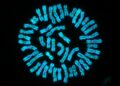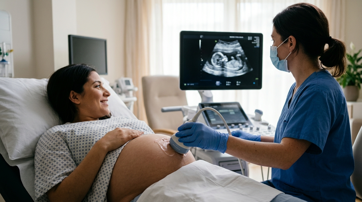
When prenatal ultrasound reveals a subtle finding that is not a structural malformation—an enlarged nuchal translucency, an echogenic bowel, a choroid plexus cyst—clinicians face a difficult question: how likely is the fetus to carry a disease-relevant genetic variant? A retrospective study from Meizhou People’s Hospital in China now offers one of the most detailed answers to date for pregnant women under 35, a group traditionally treated as low risk for chromosomal problems. Analyzing 472 fetuses whose only warning signs were these so-called non-structural ultrasound abnormalities, the researchers detected pathogenic or likely pathogenic copy number variants (P/LP CNVs) in 5.7% of cases and variants of uncertain significance (VUS) in 9.1%, according to results published in Frontiers in Genetics.
Copy number variants are deletions, duplications, insertions or complex rearrangements of genomic DNA segments longer than one kilobase. They are a major form of genomic structural variation and can disrupt embryonic development by altering gene dosage, breaking gene integrity or changing the expression of neighboring genes. Chromosomal microarray analysis (CMA) can detect these variants at far higher resolution than a conventional karyotype, which is why it has been recommended as a first-tier genetic test for fetuses with ultrasound abnormalities. Yet most existing research has focused on the general population of women referred for invasive testing, and comparatively little evidence has addressed women under 35, who are often reassured by age alone and may rely only on serological screening.
The study team, led by Lifang Lin and Liubing Lan, retrospectively enrolled 472 pregnant women aged under 35 whose fetuses showed non-structural ultrasound abnormalities and who underwent prenatal genetic testing at the hospital between October 2022 and December 2025. Inclusion required complete ultrasound and genetic data, and fetuses with definite structural malformations, multiple pregnancies, karyotype-confirmed aneuploidies or known familial chromosomal disease were excluded. The mean maternal age was 28.7 years, with a standard deviation of 3.6. Sampling followed gestational age: chorionic villus sampling at 11 to 13+6 weeks, amniocentesis at 14 to 28 weeks, and cord blood puncture after 28 weeks. Amniotic fluid samples dominated the cohort at 87.3%, with villus samples at 12.5% and a single cord blood case.
Genomic DNA extracted from these specimens was hybridized onto CytoScan 750K arrays and analyzed with the Chromosome Analysis Suite software against the GRCh37 reference genome, with quality-control thresholds set at MAPD ≤0.25 and SNP-QC ≥15. The minimum reportable size was 400 kilobases for both duplications and deletions, and the approximate lower detection limit for mosaicism was about 20%. Each CNV was classified following the 2020 American College of Medical Genetics and Genomics technical standards, weighing variant size, gene content, dosage sensitivity, population frequency and clinical databases. Importantly, the reported P/LP CNVs included only submicroscopic deletions and duplications; numerical chromosome aneuploidies were excluded by design and not counted in the P/LP statistics.
The composition of the abnormalities themselves was strikingly lopsided. Abnormal ultrasound soft markers accounted for 82.6% of cases, followed by fetal growth restriction at 12.3%, abnormal amniotic fluid volume at 4.0%, and combinations of two or more categories at just 1.1%. When detection rates were stratified by subtype, the patterns diverged between the two outcome measures. P/LP CNVs were most frequent in fetuses with abnormal amniotic fluid volume at 10.5%, followed by fetal growth restriction at 8.6%, and lowest among soft-marker cases at 4.9%. VUS showed the opposite ordering: highest in soft-marker cases at 9.2%, then fetal growth restriction at 8.6%, and lowest in abnormal amniotic fluid volume at 5.3%. None of these between-category differences reached statistical significance.
Drilling into individual soft markers, the combined P/LP-plus-VUS detection rate varied considerably, though again without statistical significance. Tricuspid regurgitation showed a combined rate of 66.7%, but this came from only 3 cases; persistent right umbilical vein and persistent left superior vena cava each showed 50.0% from just 2 cases each. The authors explicitly caution that these estimates derive from extremely small subgroups and carry substantial statistical uncertainty, and should not be read as evidence of genuinely elevated genetic risk. Among markers with more robust numbers, pyelic separation reached 25.0% (4 of 16), echogenic bowel 23.1% (3 of 13), short long bones 21.7% (5 of 23), bilateral subependymal cysts 20.0% (1 of 5), increased nuchal translucency 14.8% (19 of 128), ventriculomegaly 11.6% (8 of 69), choroid plexus cyst 10.8% (4 of 37), and nasal bone hypoplasia 9.4% (5 of 53). Aberrant subclavian artery and single umbilical artery showed no CNV-positive cases at all.
The second major question the study addressed was whether a woman’s obstetric past changes the genetic picture. Adverse obstetric history was defined as previous spontaneous abortion, induced abortion due to fetal abnormality, stillbirth, neonatal death, or delivery of offspring with congenital malformations or chromosomal disease. Of the 472 women, 368 (78.0%) had no such history and 104 (22.0%) did. The two groups were comparable in maternal age and gestational age at sampling. P/LP CNVs were found in 5.2% of fetuses from women without adverse history and 7.7% from those with it; VUS rates were 9.5% and 7.7% respectively. Neither difference was statistically significant, with p values of 0.340 and 0.701 after adjustment.
Within individual soft-marker subgroups, the comparison between women with and without adverse history also produced no significant differences after Bonferroni correction for multiple testing. One observational pattern stood out: among fetuses with echogenic bowel, the proportion of variants appeared higher when the mother had an adverse obstetric history, but this came from a subgroup of only 13 cases. The authors describe it as hypothesis-generating only, unable to support firm clinical recommendations, and call for larger independent cohorts to test it. They also note that prior research by Ronin and colleagues found the ultrasound grading of echogenic bowel was not a predictor of neonatal outcome, leaving the marker’s prognostic value unresolved.
The study’s limitations deserve careful weight. It was a single-center retrospective observational study of consecutive eligible cases, without a prospective sample-size calculation, and the observed 5.7% P/LP rate applies specifically to women under 35 referred for invasive testing because of non-structural ultrasound findings—it cannot be extrapolated to all pregnant women under 35 or to all patients with the same ultrasound features. Several soft-marker subgroups were very small, and the low number of P/LP-positive events made multivariate logistic regression statistically unreliable, so subgroup results are exploratory. The study also lacked long-term postnatal follow-up to confirm genotype-phenotype consistency, did not perform parental testing to distinguish de novo from inherited variants, and treated adverse obstetric history as a composite variable that could not be broken down by subtype. The authors further stress that the combined P/LP-plus-VUS figures used for soft-marker comparisons include VUS, which are exploratory outcomes and do not directly reflect true pathogenic genetic risk.
Despite these caveats, the clinical message is clear. Non-advanced maternal age does not eliminate the risk of fetal pathogenic CNVs when ultrasound shows non-structural abnormalities, and traditional serological screening alone is insufficient to exclude genetic risk in this group. The authors propose a tiered counseling approach: for high-risk features such as abnormal amniotic fluid volume, fetal growth restriction, and the higher-yield soft markers of pyelic separation, echogenic bowel and short long bones, CMA is strongly recommended regardless of obstetric history; for intermediate-risk isolated soft markers, CMA should be considered alongside obstetric history; and for low-risk markers such as isolated aberrant subclavian artery or single umbilical artery, routine ultrasound follow-up is preferred, with invasive testing optional after adequate counseling. For VUS results, they urge clinicians to avoid over-interpretation, fully inform families of the uncertainty, recommend serial prenatal ultrasound monitoring, and arrange standardized neonatal physical and neurodevelopmental assessment after birth. As genomic databases mature, the clinical meaning of many VUS is expected to become clearer, but for now the study reinforces that a subtle ultrasound shadow in a 29-year-old patient deserves the same genetic scrutiny it would receive in an older one.
Subject of Research: Copy number variant detection in fetuses with non-structural ultrasound abnormalities among pregnant women under 35 and its relation to adverse obstetric history
Article Title: Study on copy number variant detection results of 472 fetuses with non-structural ultrasound abnormalities in non-advanced age pregnant women and the correlation with adverse obstetric history
Article References: Lin, L., Wu, H., Zheng, Z., Chen, P., Lian, J., Huang, S., & Lan, L. (2026). Study on copy number variant detection results of 472 fetuses with non-structural ultrasound abnormalities in non-advanced age pregnant women and the correlation with adverse obstetric history. Frontiers in Genetics, 17, Article 1895948. https://doi.org/10.3389/fgene.2026.1895948
Image Credits: AI Generated
DOI: 10.3389/fgene.2026.1895948
Keywords: copy number variant, chromosomal microarray analysis, ultrasound soft markers, non-advanced maternal age, prenatal diagnosis, adverse obstetric history, variants of uncertain significance, fetal growth restriction, amniotic fluid abnormality, nuchal translucency, echogenic bowel, genetic counseling
News Source: Harold Sullivan. (November 5, 2026). Ultrasound Soft Markers in Younger Pregnant Women Flag Real CNV Risk, Study Finds. Scienmag.