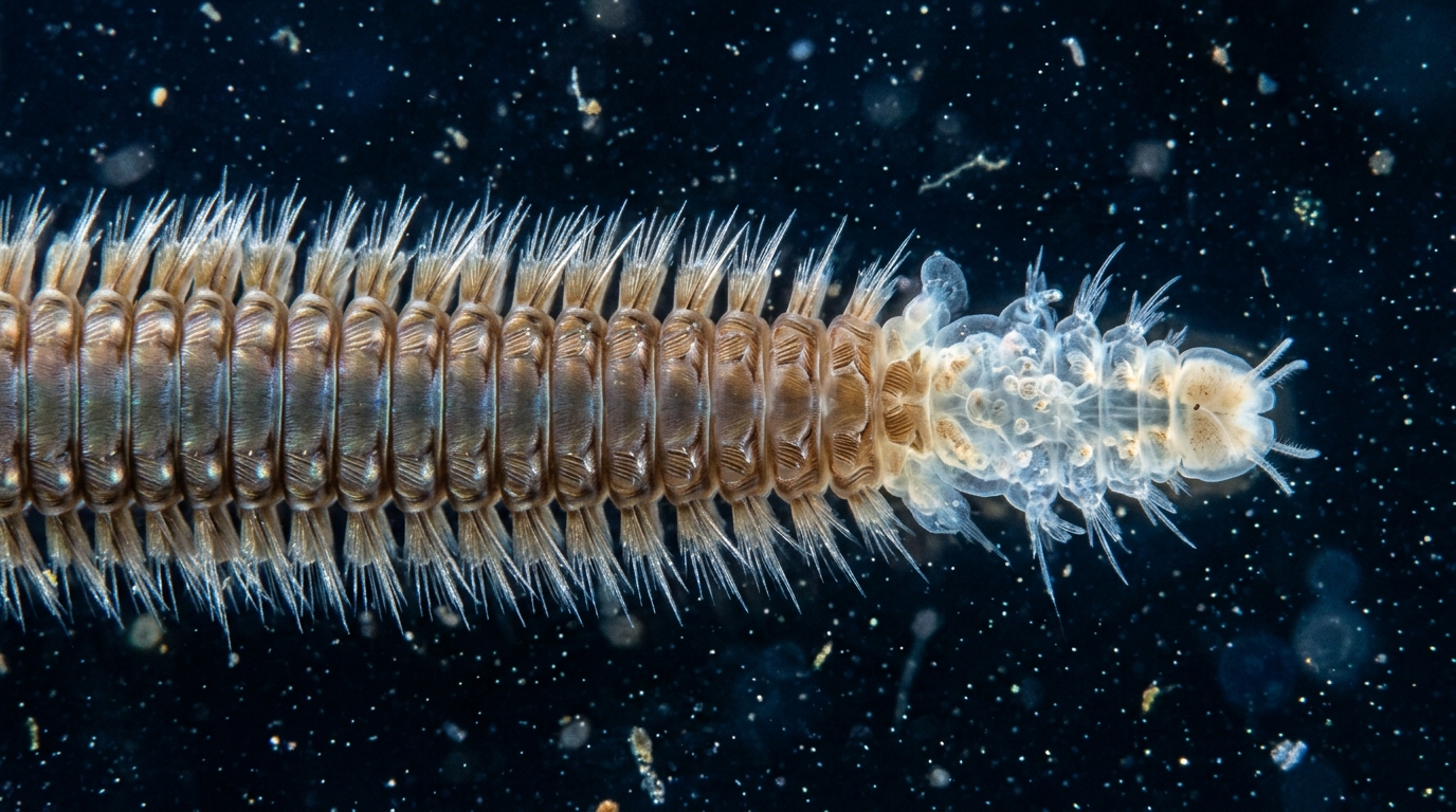
Polychaete annelids, the marine worms better known to many readers as bristle worms, carry out a feat that most animals cannot: they keep adding new body segments throughout their lives, and they can rebuild lost segments after amputation. A new review published in Development Genes and Evolution by Benoni Boilly, Hubert Hondermarck and M. Teresa Aguado pulls together decades of experimental work to explain what makes this possible. The authors conclude that two ingredients are indispensable for the segment-building machinery to operate: a supply of mesodermal regeneration cells and the presence of regenerating nerve fibres near the site of segment production.
The engine of both normal growth and segment regeneration is a narrow band of tissue called the segment addition zone, or SAZ. In intact worms, this zone sits just in front of the pygidium, the posterior-most body end, and continuously generates new segments that differentiate in a postero-anterior direction, lengthening the animal. When posterior segments are amputated, the worm first regenerates a pygidium, and a new SAZ then forms adjacent to it and produces replacement segments. The SAZ is described as a thin transversal region made of two rings of proliferating cells, one ectodermal and one mesodermal, and its output follows a characteristic S-shaped growth curve: fast at first, then slower as regeneration shades into ordinary growth.
The review’s central evidence comes from experiments performed during the 1960s and 1970s on three polychaete groups: Syllis amica, Nereis diversicolor, now known as Hediste diversicolor, and Aricia foetida, currently Phylo foetida. When whole worms were X-irradiated before or soon after amputation, no segments regenerated. More telling were the selective experiments. Researchers destroyed the mesodermal regeneration cells completely, either with X-rays or by microinjecting the coelomic cavity with Thorotrast, a radioactive suspension of thorium dioxide. Cytological examination confirmed that these treatments hit coelomic epithelial cells while leaving epidermal and intestinal epithelia intact.
The outcome was strikingly specific. Worms deprived of their mesodermal regeneration cells still regenerated a pygidium after posterior amputation, and in species capable of anterior regeneration they still rebuilt a prostomium, the head end. What they could not do was make new segments. The SAZ simply failed to function. With incomplete destruction of the mesodermal cells, some segments did regenerate, and their number fell in proportion to the X-ray dose or Thorotrast concentration used. The authors interpret this as showing that the extent of segment regeneration depends on how many viable mesodermal blastema cells survive the treatment.
In Syllis amica, all blastema cells originate from the amputated segment itself, without migration between segments, each tissue compartment of the blastema arising from the corresponding stump tissue through partial dedifferentiation followed by redifferentiation. The last segment of the stump supplies the blastema’s mesodermal cells, while the posterior stump epidermis and intestinal epithelium yield the epidermal and gut cells of the regenerate. Activated mesodermal cells show a distinctive cytological profile: a large nucleus, a prominent nucleolus surrounded by a clear zone, and strongly basophilic cytoplasm, consistent with high rRNA synthesis. The authors note that these cells’ high sensitivity to X-irradiation may relate to their low differentiation and high proliferative activity.
The second indispensable factor is nervous. After segment amputation, nerve fibres proliferate rapidly from the cut nerve cord and creep between the epidermis and the mesodermal regeneration cells toward the regenerated pygidium, where the mesodermal cells are well activated. The review argues that these fibres, and the growth factors they secrete, sustain the proliferation of the regeneration cells. Direct proof is difficult because the nerve cord cannot be selectively destroyed, but natural double nerve cords in some Nereis specimens offered a clue: each cord corresponded to a normal tail, occasionally producing two tails from the same segment.
Anterior regeneration reveals the nervous system’s role from a different angle. In syllid worms that can regenerate a head, nerve fibres grow from the severed cord toward the differentiating brain of the new prostomium. This draws the regenerating fibres away from the amputation plane, where the transient SAZ of anterior regeneration sits, leaving it relatively denervated. The authors propose that this loss of local nerve contact, and of the growth factors the fibres provide, explains why anterior segment production is so limited. In Syllis amica, anterior regeneration produces only three to four segments and stops by about 45 days post-amputation, whereas posterior regeneration yields around 23 segments by 60 to 70 days and then grades into normal growth.
The digestive tube adds a further layer of difference. In posterior regeneration, the gut is necessary for segmentation; worms deprived of the digestive tube by microdissection or Thorotrast poisoning lost SAZ function. The coelomic epithelium surrounding the intestine is a major source of mesodermal regeneration cells, and the authors suggest this supply is what anterior regeneration in S. amica lacks, since the worm does not rebuild its intestine forward. Supporting this, two related syllids, Syllis gracilis and Syllis malaquini, which can regenerate the entire stomodeal anterior digestive tube, produce far more anterior segments. The authors also report that decerebrated nereid worms can regenerate a pygidium but fail at segmentation even with a normal nerve cord, an observation to be read cautiously because brain removal triggers sexual maturation and spawning-related transformations that divert energy away from regeneration.
The authors checked whether sexual maturation or stolonization could explain the weakness of anterior regeneration by re-amputating posterior segments 50 and 75 days after the first cut, with or without X-irradiation and even after stolon release. All still regenerated three to four anterior segments on the same schedule, arguing against that confound. They also point to recent transcriptomic work comparing limited and strong anterior regenerators among syllids, which found that gene expression during posterior regeneration and normal growth is similar but not identical to that of anterior regeneration, though which genes drive the different phases remains open.
What emerges is a two-factor model. The terminal structures, pygidium and prostomium, regenerate independently of mesodermal cells, echoing the two halves of the trochophore larva before its mesodermal primordia appear. Segment formation, by contrast, requires both mesodermal regeneration cells and sustained nerve-derived stimulation. Posterior regeneration enjoys both: the gut’s coelomic epithelium supplies cells, and nerve fibres remain in contact with the SAZ throughout regeneration and normal growth. Anterior regeneration in S. amica has neither advantage. The authors stress that the precise nature of the nerve factor, and the lineage of the mesodermal blastema cells, still need molecular-level investigation, with single-cell transcriptomics in more closely related species flagged as a promising route. They even propose a testable experiment: keeping the nerve fibres of an anteriorly regenerating worm open by microsurgery or laser ablation to see whether anterior segment production improves.
Subject of Research: The roles of mesodermal regeneration cells and nerves in segment regeneration of polychaete annelids
Article Title: Nerves and availability of mesodermal cells are essential for the function of the segment addition zone (SAZ) during segment regeneration in polychaete annelids
Article References: Boilly, B., Hondermarck, H., & Aguado, M. T. (2024). Nerves and availability of mesodermal cells are essential for the function of the segment addition zone (SAZ) during segment regeneration in polychaete annelids. Development Genes and Evolution, 234(2), 65-75. https://doi.org/10.1007/s00427-024-00713-5
Image Credits: AI Generated
DOI: 10.1007/s00427-024-00713-5
Keywords: polychaete annelids, segment addition zone, posterior regeneration, anterior regeneration, mesodermal regeneration cells, nerve cord, growth factors, pygidium, prostomium, Syllis amica, X-irradiation, Thorotrast
News Source: Drew Townsend. (November 5, 2026). Nerves and Mesodermal Cells Drive Segment Regeneration in Polychaete Annelids. Scienmag.