


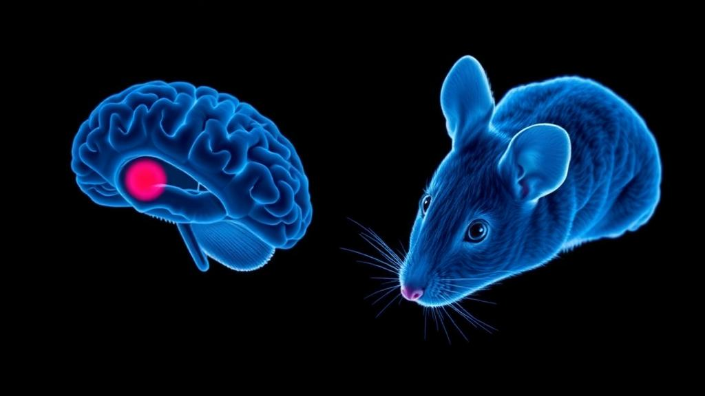
Researchers at Stanford University report a way to activate gene editing at selected locations deep in the body using focused ultrasound and light-emitting nanoparticles. In an experimental study in mice, the team used ultrasound to trigger nanoparticles near a targeted site to emit light. That light switched on a light-sensitive CRISPR-family editor, which could then act on a selected gene. When the researchers focused ultrasound on one side of a mouse’s brain, that side began to glow while the other remained dark, indicating that gene disruption had been spatially controlled in the tested model.
The approach addresses two related questions: which gene an editor can affect and where in the body that activity occurs. CRISPR tools can be designed to act on particular genes, but selecting a gene does not itself restrict editing to one location. The release describes this as a limitation for possible efforts to affect chronic pain in one region without changing sensation elsewhere. In the researchers’ example, gene choice alone would not supply that spatial selectivity. A technique that also selects a location could, in principle, let researchers ask where a gene should be altered, as well as which gene to target. The release notes that localized delivery is possible, but describes implanted nanoscaffolds as one invasive approach. Such scaffolds release CRISPR tools locally and affect only a handful of cells, according to the release. This example illustrates both the local reach and the invasive nature of that option; it does not establish how all localized delivery methods perform. The Stanford team sought a way to select a site without surgical implants. Their proposed arrangement makes the location choice through focused ultrasound: the focus identifies where nearby nanoparticles emit light, while the light-sensitive editor responds to that illumination. In this design, spatial control depends on triggering the switch at the selected site, while the editor itself is designed for a particular gene. This is the rationale for combining the components, rather than treating gene specificity as sufficient to determine where editing happens. The report describes an experimental mouse demonstration of this strategy. It does not establish that it solves the targeting problem in people, works across all tissues, or provides a pain treatment. The pain example remains a motivation for spatial control, while the reported experiment concerns location-specific editing in mice.
Light can control engineered proteins and cellular processes, and light-sensitive gene editors can be switched on by illumination. But light does not travel far through biological tissue, making it difficult to reach deep targets from outside the body. Focused ultrasound penetrates tissue more deeply. Guosong Hong’s group had developed nanoparticles that circulate in the bloodstream and emit light when struck by focused ultrasound; Michael Lin’s group had been engineering CRISPR-Cas9 editors that respond to light. Combining the two offered a route to activate editing at an ultrasound-selected location, provided the light source and editor could work together in tissue.
Each component had a constraint. Lin’s existing light-sensitive editor was too large for a single viral delivery vehicle, described as the usual way to get CRISPR molecules into cells. Hong’s nanoparticles also needed to produce enough light without requiring potentially tissue-damaging ultrasound intensity. The groups modified both. Postdoctoral scholar Daesun Song developed a smaller Cas12f-based light-sensitive editor and engineered it to respond more efficiently to light. Hong’s group made the nanoparticles brighter, reducing the ultrasound power needed to activate the editor, according to the release. These changes addressed the reported delivery-size and light-generation challenges. The account gives no numerical size or activation threshold and does not quantify tissue safety or define a safe operating range. Spatial editing still depends on delivering the editor, focusing ultrasound, and generating light at the selected site. The report does not say either modification alone provides spatial editing. It describes the engineering rationale and use of the combined system in mice, not a performance guarantee for other editors, particles, or tissues. The release reports the linked design steps but does not quantify their individual effects or establish how the system performs in other settings.
In the reported sequence, the researchers first delivered a Cas12f editor designed to target a particular gene. They then focused ultrasound at a chosen location. Sound waves caused nearby nanoparticles to emit light, and the light activated the editor so it could act on that gene. The method joins three functions: the editor supplies the gene target, the ultrasound focus selects a region, and the light-sensitive switch links the two by limiting when the editor is active. The release describes the work as noninvasive and reports no surgery or implanted device as part of the demonstration.
For a spatial readout, researchers used a mouse modified so tissues glowed but a second genetic change suppressed that signal. Disrupting the stop signal would allow glow. After ultrasound was directed at one side of the brain, that side glowed while the other remained dark, showing location-specific gene disruption in this model. The signal indicates where the targeted disruption occurred; it is not a measure of symptoms, behavior, or therapeutic benefit. The account does not show that every cell on the targeted side was edited or resolve activity at finer scales. This experiment demonstrates localized gene disruption in an animal using ultrasound, light-emitting nanoparticles, and a light-sensitive editor. It does not establish performance in other brain regions or tissues, safety for disease treatment, or clinical benefit. The mouse’s prior modifications enabled the visible reporter outcome, supporting the limited conclusion that editing location was controlled in this model. The reported difference is between the targeted and untargeted sides; the release does not provide a cell-by-cell distribution or measure how precisely activity was confined within either side.
The release points to neuropathic pain as one possible future application. Research in mice suggests that silencing genes associated with sodium ion channels using CRISPR techniques can control neuropathic pain, but the approach tends to numb the entire body. The researchers suggest that silencing those genes only in the part of the nervous system associated with pain might eventually permit more targeted relief. That possibility remains prospective: the reported experiment tested spatial gene editing in mouse brains, and the release does not report a pain-treatment trial using this system. The same ability to target different brain regions could also help researchers investigate how genes function in distinct areas.
The work was published in Cell on September 28, 2026, under the title “Non-invasive control of gene editing in vivo by photoswitchable Cas12f and focused ultrasound.” The release identifies it as an experimental study involving animals. It describes the mouse-brain demonstration, alongside ongoing work in the two research groups that may extend the approach. Lin’s group is working on a variation intended to temporarily activate or repress genes without changing the underlying DNA. This is a different proposed function from editing a gene by altering DNA, and the release presents it as work in progress rather than an outcome of the reported experiment. Hong’s group is developing a wearable ultrasound device intended to let researchers control genes while an animal moves and observe behavioral effects. The device is described as being developed; the release does not say that it was used in the reported brain experiment or that it has already enabled such observations. These directions relate to the same broader interest in controlling gene activity at selected locations, but they remain distinct from the published demonstration. The article tested spatial gene disruption in a mouse using focused ultrasound, light-emitting nanoparticles, and a photoswitchable Cas12f editor. The follow-on efforts describe possible changes to the type of gene control and to the setting in which researchers might apply ultrasound. Neither is evidence that the method has produced behavioral findings or temporary gene regulation. The report also does not establish clinical use, and its release frames future clinical relevance as a possibility. Keeping the findings and development plans separate clarifies what is known from this study: a selected brain side showed the glow readout after ultrasound targeting, while the other remained dark. The other directions describe ongoing research, not results established by that animal experiment.
The system’s central contribution, as presented by the researchers, is combining a light-sensitive editor with a remotely triggered light source so that gene editing can be controlled by location as well as by gene choice. The reported glow on only the ultrasound-targeted side of the brain serves as the spatial readout in the demonstration. The release does not establish how the method performs across different tissues, genes, or treatment settings, nor does it show that it is ready for use in patients. Its proposed applications, including localized pain relief, remain future possibilities while the team develops and tests the technology.
Subject of Research: Ultrasound-triggered, light-sensitive gene editing in mice
Article Title: New technology controls gene editing noninvasively with ultrasound and light
Article References: New technology controls gene editing noninvasively with ultrasound and light. Original publication (Bibliographic details unavailable.)
Image Credits: AI Generated
DOI: Not provided
Keywords: gene editing, CRISPR, ultrasound, nanoparticles, Cas12f, mouse study, neuroscience, biotechnology, technology, controls, gene, editing
News Source: Juliet Wilcox. (October 29, 2026). Ultrasound and Light Guide Gene Editing to Selected Sites in Mouse Brains. Scienmag.



