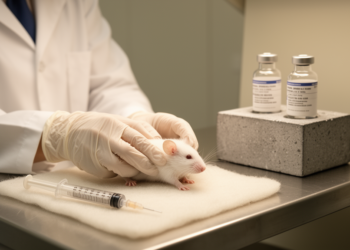
Testosterone replacement therapy may help or harm the brain in Parkinson’s disease depending on when treatment begins, according to a narrative review published in Discover Neuroscience that surveys the current preclinical and clinical evidence. The review, led by José Leandro S. Souza and colleagues, concludes that the therapeutic hypothesis is biologically plausible but not yet recommendable for clinical treatment, because the clinical literature is too small and heterogeneous to define a therapeutic window and no systematic dose-finding study supports a Parkinson’s-specific dosing recommendation.
Testosterone is a steroid hormone synthesized from cholesterol, produced primarily by Leydig cells in the testes in men, while in women it derives from androstenedione made by the adrenal cortex and theca cells, with women typically producing 20 to 30 times less than men. Its synthesis is regulated through negative feedback involving hypothalamic neurons that release gonadotropin-releasing hormone, which stimulates the anterior pituitary to secrete follicle-stimulating hormone and luteinizing hormone; luteinizing hormone acts on Leydig cells to promote testosterone synthesis and release. The hormone performs critical physiological functions, including maintaining sexual health, promoting muscle mass and bone density, and regulating the cardiovascular system, metabolic processes, liver function, the immune system, and the central nervous system. Endogenous production declines progressively with age, beginning around the fourth decade of life.
At the cellular level, testosterone’s lipophilic nature allows it to diffuse across cell membranes and bind to androgen receptors in the cytoplasm or nucleus, forming a hormone-receptor complex that interacts with hormone response elements in DNA to initiate gene transcription and new protein synthesis over hours or days, the so-called genomic mechanisms. Non-genomic mechanisms involve binding to membrane-associated receptors, including G protein-coupled receptors and tyrosine kinase-associated receptors, triggering rapid second messenger cascades through pathways such as PKA/PKC, PI3K/AKT, and MAPK. In the nervous system, androgen receptors are expressed in various regions, including dopaminergic neurons, and testosterone has been shown to upregulate genes encoding tyrosine hydroxylase, catechol-O-methyltransferase, monoamine oxidases A and B, vesicular monoamine transporter 2, the dopamine transporter, and dopamine receptors D1, D2, and D5, effects accompanied by increased dopamine concentrations in the striatum.
Parkinson’s disease is the second most prevalent neurodegenerative disorder worldwide, a progressive condition marked primarily by degeneration of dopaminergic neurons in the substantia nigra pars compacta and, to a lesser extent, the ventral tegmental area. The neuronal loss reduces dopaminergic projections and dopamine availability in the dorsal striatum, disrupting basal ganglia circuitry and producing hallmark motor symptoms such as bradykinesia. The primary histopathological hallmark is the accumulation of misfolded proteins forming Lewy bodies, composed predominantly of alpha-synuclein, whose aggregation is strongly associated with elevated oxidative stress, mitochondrial dysfunction, and neuroinflammatory responses. Non-motor symptoms, including sleep disorders, autonomic dysfunction, anxiety, depression, cognitive impairment, and apathy, often precede motor manifestations, and patients often present with changes in body mass, sarcopenia, and reduced circulating testosterone concentrations.
Recent studies have demonstrated that patients with Parkinson’s disease exhibit significantly reduced plasma testosterone levels compared to age-matched healthy controls. However, the review stresses that with the scarcity of studies to date, it cannot be stated that low testosterone levels serve as a biomarker of disease severity, a consequence of the disease, or a factor contributing to progression, although the findings support an interaction between androgen signaling and pathology. The authors also caution that the age-related drop in testosterone should not, in and of itself, be interpreted as an indication for replacement therapy or as evidence that testosterone deficiency causes Parkinson’s disease. The disease shows a pronounced gender disparity, with higher prevalence and more rapid progression in males, a difference that has been linked to sex hormones.
The preclinical evidence reveals a striking duality. In one study, a 12-week regimen of testosterone propionate at 1 milligram per kilogram per day in aged male rats enhanced mitochondrial function, increasing mitochondrial membrane potential and complex V activity, upregulated mitochondrial biogenesis markers including PGC-1α, NRF-1, and TFAM, boosted endogenous antioxidant enzymes, and preserved neuronal populations in the substantia nigra and hippocampus while preventing age-associated cognitive and behavioral impairments. Similar administration in aged rats increased NRF-2 and PGC-1α expression, raised dopaminergic markers including tyrosine hydroxylase and the dopamine transporter, and increased striatal dopamine content. Chronic testosterone propionate at 1 milligram per kilogram per day also attenuated the loss of tyrosine hydroxylase-positive neurons in a reserpine-induced model of parkinsonism, preserving striatal immunoreactivity and improving motor performance.
Further supporting evidence comes from castrated mice, which developed molecular and behavioral phenotypes characteristic of Parkinson’s disease, with increased neuroinflammatory and glial activation markers such as GFAP, Iba-1, iNOS, IL-1β, and TNF-α in the substantia nigra, pathological alpha-synuclein accumulation, dopaminergic cell loss, and striatal denervation. Daily administration of 15 milligrams of dihydrotestosterone for 30 consecutive days immediately after castration preserved striatal dopamine levels and prevented significant motor impairments. In a progressive model using repeated low-dose reserpine, low-dose testosterone treatment at 1 milligram per kilogram preserved tyrosine hydroxylase-positive neurons in the substantia nigra, ventral tegmental area, and striatum and significantly improved motor impairments, whereas a higher dose of 5 milligrams per kilogram showed limited effects, suggesting the neuroprotective effect depends on dose. Work in elderly Nrf2-knockout mice indicates the Nrf2 pathway appears to be a central mechanism of testosterone-induced neuroprotection, since the benefits on mitochondrial function, PGC-1α expression, and tyrosine hydroxylase preservation practically disappear when Nrf2 is absent.
Yet the hormone can aggravate pathology under other conditions. Elevated testosterone levels have been linked to neurotoxicity, particularly under heightened oxidative stress, where they may exacerbate the loss of dopaminergic neurons. In cultured dopaminergic neurons subjected to hydrogen peroxide-induced oxidative stress, testosterone administration exacerbated inflammation, marked by increased expression of NF-κB and cyclooxygenase-2, and significantly reduced cell viability compared to untreated controls. In aged rats pretreated with reserpine, testosterone amplified lipid peroxidation, decreased mRNA expression of Nrf2, heme oxygenase-1, and NQO1, reduced levels of tyrosine hydroxylase, the dopamine transporter, and dopamine in the nigrostriatal pathway, and produced pronounced motor impairments, while in animals not subjected to prior reserpine-induced damage it exerted neuroprotective effects. The review concludes that neuroprotective effects emerge primarily under conditions of chronic, progressive neurodegeneration or age-related androgen decline, whereas neurotoxic effects appear predominantly in settings of acute oxidative stress and severe pre-existing neuronal injury, making the neurobiological context, rather than testosterone itself, the likely major determinant of outcome.
The clinical picture remains far less settled. The first clinical study involved only 10 male participants and reported improvements in both motor and non-motor symptoms following daily transdermal testosterone gel, which provided relatively stable plasma levels. A later study of 30 men, 15 of whom received replacement therapy, found no significant differences in motor or non-motor outcomes between treatment and control groups; the earlier trial used a daily transdermal gel, while the later one employed intramuscular injections of testosterone enanthate every two weeks, a regimen that, given the approximately 4.5-day half-life of testosterone enanthate, likely produced substantial fluctuations in plasma concentrations. Notably, patients who applied topical testosterone gel daily for one month reported reduced sleepiness, increased energy levels, greater life satisfaction, and improved work performance, but the subsequent eight-week intramuscular regimen of 200 milligrams every two weeks did not yield significant improvements. More recently, a case-control study involving 923 participants found that elderly men undergoing testosterone replacement therapy exhibited a significantly reduced risk of developing Parkinson’s disease, though the authors note that therapeutic outcomes remain inconsistent across studies.
Translating these findings into treatment faces substantial obstacles. No studies conducted in people with Parkinson’s disease, or in animals, provide a consistent relationship between therapy exposure and an effective clinical response, and the absence of dose-response studies means experimental doses cannot be extrapolated directly to patients. Most safety evidence originates from non-PD populations; potential adverse effects include erythrocytosis, fluid retention, hypertension, acne, gynecomastia, infertility, and worsening of obstructive sleep apnea, and treatment should be avoided in men with confirmed or suspected prostate or breast cancer, elevated hematocrit, obstructive sleep apnea, severe lower urinary tract symptoms, heart failure, myocardial infarction or recent stroke, and thrombophilia, contraindications drawn mainly from the hypogonadism literature rather than Parkinson’s-specific trials. The clinical studies to date suffered from small sample sizes, short follow-up periods, and heterogeneous protocols, and because this is a narrative review without formal assessment of study quality or risk of bias, selection bias cannot be excluded. The authors propose that future trials enroll clearly defined hypogonadal populations confirmed by repeated morning testosterone measurements, stratify by disease stage, compare stable exposure regimens, measure serum testosterone longitudinally, and record motor and non-motor outcomes, body composition, cardiovascular, sleep, and prostate health, before testosterone therapy can be considered a disease-modifying option for Parkinson’s disease.
Subject of Research: The potential benefits, risks, and clinical limitations of testosterone replacement therapy in Parkinson's disease
Article Title: Testosterone replacement therapy in Parkinson’s disease a narrative review of current evidence
Article References: Souza, J. L. S., Rocha, G. S., Bispo, J. M. M., Luz, A. C. A., Silva, R. H., Ribeiro, A. M., Medeiros, K. A. A. L., Gois, A. M., Freire, M. A. M., & Santos, J. R. (2026). Testosterone replacement therapy in Parkinson’s disease a narrative review of current evidence. Discover Neuroscience, 21(1), Article 104. https://doi.org/10.1186/s13064-026-00335-9
Image Credits: AI Generated
DOI: 10.1186/s13064-026-00335-9
Keywords: testosterone replacement therapy, Parkinson's disease, narrative review, neuroprotection, dopaminergic neurons, substantia nigra, oxidative stress, neuroinflammation, preclinical studies, clinical trials, androgen signaling, aging
News Source: Cassandra Pierce. (October 29, 2026). Testosterone Therapy Shows Mixed Effects in Parkinson’s Disease, Review Finds. Scienmag.