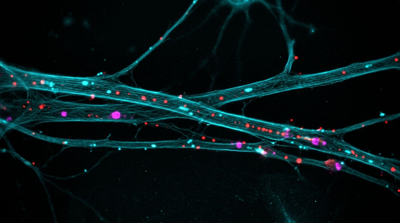
Pathological forms of the RNA-binding protein TDP-43 sabotage a tankyrase-driven control system that neurons rely on to keep their proteasomes assembled, active, and moving along axons, according to a study published in the Journal of Biomedical Science. The work, led by Pitchapa Bubphachat, Pritha Majumder, and colleagues with senior author Chi-Chen Sarah Huang, combined cell culture models, live-cell imaging, and a transgenic mouse line carrying a familial ALS-linked TDP-43 mutation to map how the protein disrupts protein quality control in neurons. The findings identify the TNKS/PI31/proteasome axis as a mechanistically relevant pathway linking pathological TDP-43 to proteasome dysfunction and impaired axonal proteostasis, and they suggest that selectively restoring this pathway may offer a more precise therapeutic strategy than broadly activating the proteasome.
The stakes are high because TDP-43 pathology sits at the center of two devastating diseases. Ubiquitinated inclusions are the pathological hallmark of amyotrophic lateral sclerosis and frontotemporal lobar degeneration, and TDP-43-positive ubiquitinated inclusions constitute the major pathological component of these inclusions in 95 percent of ALS patients and 45 percent of patients with frontotemporal lobar degeneration with ubiquitin-positive inclusions. Under physiological conditions, TDP-43 is predominantly nuclear, where it regulates RNA processing and metabolism, while a smaller cytoplasmic pool contributes to mRNA stability, transport, and translation. In disease, the protein becomes hyperphosphorylated, polyubiquitylated, cleaved into C-terminal fragments, and sequestered into detergent-insoluble cytosolic aggregates in the hippocampus, cortex, and spinal cord motor neurons. Multiple independent clinical studies have also reported a 1.2 to 1.5-fold increase in total TDP-43 protein in the brain tissue, plasma, and cerebrospinal fluid of ALS and FTLD patients compared with healthy controls, and the study authors note that this slight increase is sufficient to promote pathological misfolding.
To mimic that disease state, the researchers overexpressed GFP-tagged human wild-type TDP-43 full-length, the A315T mutant, or the truncated TDP-25 fragment in mouse neuroblastoma N2a cells, using equal amounts of plasmid DNA in equal numbers of cells. Sequential RIPA and urea fractionation, semi-denaturing detergent agarose gel electrophoresis, and immunofluorescence revealed distinct aggregation patterns. GFP alone remained soluble and diffuse, but wild-type full-length TDP-43 and the A315T mutant preferentially accumulated in the urea-insoluble fraction as oligomeric species while remaining mainly nuclear without prominent cytoplasmic inclusions. TDP-25 behaved differently, showing reduced RIPA-soluble levels, predominant insoluble oligomeric accumulation, and prominent perinuclear and cytoplasmic punctate aggregates. Despite these differences in aggregation propensity, all three pathological species reduced endogenous 20S proteasome activity, measured with the fluorogenic substrate Suc-LLVY-AMC, with the strongest inhibition seen in cells expressing the A315T mutant or TDP-25. The proteasome and loading control levels were comparable across groups, ruling out differences in proteasome abundance or protein loading as confounders.
Functional consequences followed. The researchers used a GFP-tagged ubiquitin mutant substrate, UbG76V-GFP, whose expression is inversely correlated with intracellular 26S proteasome activity, and found significantly increased UbG76V-GFP levels in NSC34 cells overexpressing TDP-43-FL or TDP-25, with the most pronounced accumulation in TDP-25-expressing cells. Cells expressing any of the three pathological TDP-43 constructs also showed significant increases in endogenous polyubiquitylated protein levels compared with the GFP control group. In parallel, the team verified the effect in tissue from Prp-TDP-43A315T transgenic mice, which express Flag-tagged human TDP-43-A315T under the mouse Prp promoter and develop a progressive, fatal neurodegenerative phenotype resembling both ALS and FTLD-U. Although TDP-43 aggregates were not observed in the transgenic spinal cord, phosphorylated TDP-43 oligomers and polymers were significantly detected during disease progression, with the highest levels at late stages. Twenty S proteasome activity was significantly reduced in transgenic spinal cord compared with wild-type littermates, and the activity-based probe MV151, which covalently labels the catalytic beta-1, beta-2, and beta-5 subunits of active 20S proteasomes, confirmed reduced activity in both the transgenic tissue and the transfected cells, with no signal in MG132-treated controls.
The study then moved to a question that had not been addressed before: whether TDP-43 pathology disrupts the transport of proteasomes along axons, a process essential for local protein quality control in neurons with long axons. Using time-lapse live-cell imaging of MV151-labeled 20S proteasomes in primary cortical neurons from wild-type E16.5 mouse brains, the researchers found that the percentage of moving proteasomes was significantly reduced in neurons transfected with GFP-TDP-25 compared with GFP controls. The labeled proteasomes in TDP-25-expressing neurons also showed significant decreases in average velocity and net displacement, and a comparable impairment was observed in neurons expressing full-length TDP-43. Proteasomes moving faster than 0.1 micrometers per second within a 10 to 20 second tracking window were classified as moving, while slower particles were considered stationary, and the analysis relied on pooled technical measurements from three independent biological replicates.
To explain these defects, the investigators focused on tankyrase 1 and 2, poly ADP-ribose polymerases that ADP-ribosylate the proteasome inhibitor PI31. Under physiological conditions, this modification releases PI31 from the 20S core, relieving its inhibitory grip and allowing the 19S regulatory particle to assemble with the 20S catalytic core into the active 26S proteasome. PI31 also serves as an adaptor for axonal transport, directly binding dynein light chain LC8-type proteins to promote fast axonal transport of proteasomes between the soma and neuron terminals. Förster resonance energy transfer assays in primary hippocampal neurons showed that TNKS interacts with both wild-type TDP-43 and the A315T mutant in the cytoplasm, and with PI31 as well. Immunoprecipitation from transgenic mouse spinal cord lysates then revealed lower TNKS-PI31 association and attenuated PI31 ADP-ribosylation compared with wild-type controls, indicating that pathological TDP-43 is associated with reduced TNKS-PI31 interaction and TNKS-mediated PI31 modification.
Fractionation experiments pointed to sequestration as the mechanism. Levels of insoluble TNKS and 19S proteasome subunits, including PSMC6 and PSMD4, in the urea fraction were significantly higher in transgenic spinal cord than in wild-type controls, and endogenous TNKS showed significantly increased colocalization with pathological TDP-43 in transgenic primary motor neurons. Native gel electrophoresis showed reduced levels of structurally intact 26S, 20S, and 19S proteasome complexes and an increased ratio of disassembled 19S and 20S subcomplexes to intact 26S complexes in the transgenic mice, alongside attenuated in-gel 20S proteasome activity. The proportion of TDP-43 colocalized with MV151-labeled 20S proteasomes was also significantly increased in transgenic motor neurons. Collectively, the authors conclude, pathological TDP-43 sequesters TNKS and a subset of proteasome components into insoluble fractions, reducing TNKS availability for interaction with PI31 and limiting the availability of soluble 19S and 20S subcomplexes for 26S proteasome assembly.
A key experiment tested whether the TDP-43-TNKS interaction itself drives the damage. The team generated a TDP-43 mutant, TDP-43-TBD, carrying H166A and I168A substitutions within the tankyrase-binding motif in the RRM1 domain. This mutant aggregated even more aggressively than full-length TDP-43, accumulating predominantly in the insoluble urea fraction and forming pronounced cytoplasmic aggregates, yet it sequestered far less TNKS into the insoluble fraction, showed markedly reduced interaction with TNKS, and permitted increased TNKS-PI31 binding. Critically, even though TDP-43-TBD was more prone to form insoluble aggregates than TDP-43-A315T, it did not impair 20S proteasome activity as significantly as the A315T mutant did. The authors conclude that aggregation of TDP-43 alone is insufficient to disrupt proteasome function; instead, the interaction between TDP-43 and TNKS is an important contributor to TNKS sequestration, impairment of the TNKS/PI31 signaling axis, and subsequent proteasome inactivation.
Pharmacological inhibition of TNKS with XAV939, a cell-permeable selective inhibitor that blocks TNKS-mediated PI31 ADP-ribosylation, independently confirmed the pathway. In wild-type primary cortical neurons, XAV939 significantly reduced PI31 ADP-ribosylation, increased PI31 binding to the 20S proteasome, and reduced 26S proteasome activity, recapitulating the effects observed in neurons from the transgenic mice. In live primary motor neurons derived from E12.5 embryos, around 60 percent of cells were CHAT-positive motor neurons, and time-lapse imaging showed that proteasomes in transgenic mice were less dynamic than those in wild-type controls, with significant decreases in average velocity and net displacement in both retrograde and anterograde directions. XAV939 treatment reproduced these deficits in wild-type neurons as well: the percentage of moving 20S proteasomes fell from 57 percent in DMSO-treated wild-type controls to 43 percent with XAV939, and to 38 percent in XAV939-treated transgenic neurons compared with 36 percent in DMSO-treated transgenic neurons. The proportion of retrograde movement rose from 47 percent in DMSO-treated wild-type neurons to 68 percent in XAV939-treated transgenic neurons, a shift the authors interpret cautiously, noting that because absolute retrograde flux and proteasome delivery to the soma were not directly measured, this may reflect a compensatory response to proteotoxic stress, though alternative explanations such as impaired anterograde transport cannot be excluded.
The spatial consequences were equally striking. In transgenic motor neurons, TDP-43 accumulated mainly in the cell body and proximal axons while being reduced or almost absent in distal axons, and MV151-labeled 20S proteasomes and PI31 were predominantly clustered in the proximal axonal region, 0 to 50 micrometers from the cell body, while rarely appearing in the distal region, 50 to 80 micrometers from the cell body. The proportion of PI31 colocalized with MV151-labeled 20S proteasomes in the axon was significantly increased in transgenic motor neurons, and XAV939 treatment reproduced this proximal accumulation and increased colocalization in both wild-type and transgenic neurons. The authors acknowledge that their cellular models and the Prp-TDP-43A315T transgenic mice cannot fully recapitulate the genetic, cellular, and pathological heterogeneity of human ALS and FTLD, and they call for validation in patient-derived neuronal systems and future studies using genetic approaches and patient-derived iPSC motor neurons. Within those limits, the study identifies a functional TNKS/PI31/20S proteasome axis that contributes to the regulation of proteasome activity and axonal trafficking, and it proposes that blocking the aberrant binding between TNKS and TDP-43, rather than indiscriminately activating the proteasome, may offer a targeted way to restore proteasome function and ameliorate the pathological hallmarks of TDP-43 proteinopathies while minimizing off-target effects.
Subject of Research: How pathological TDP-43 disrupts the TNKS/PI31/proteasome axis in ALS and frontotemporal lobar degeneration models
Article Title: The TNKS/PI31–proteasome axis as a central node in TDP-43 neuropathies
Article References: Bubphachat, P., Majumder, P., Wang, I. F., Huang, J. K., Kao, T.-J., Lee, Y.-C., Shen, C.-K. J., Chen, G.-C., Chen, P.-C., & Huang, C.-C. S. (2026). The TNKS/PI31–proteasome axis as a central node in TDP-43 neuropathies. Journal of Biomedical Science, 33(1), Article 94. https://doi.org/10.1186/s12929-026-01296-1
Image Credits: AI Generated
DOI: 10.1186/s12929-026-01296-1
Keywords: TDP-43, ALS, frontotemporal lobar degeneration, proteasome, tankyrase, PI31, axonal transport, protein homeostasis, XAV939, ADP-ribosylation, Prp-TDP-43A315T mice, neurodegeneration
News Source: Ophelia Keating. (November 6, 2026). TDP-43 Hijacks a Tankyrase Switch That Powers Proteasome Assembly and Axonal Transport. Scienmag.