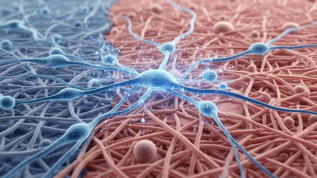
A high-resolution transcriptomic investigation has revealed that the divergent clinical behaviors of keloid scars correspond to distinct cellular trajectories and specialized vascular programs within endothelial cells. Keloids represent benign yet locally invasive fibroproliferative skin disorders characterized by excessive extracellular matrix deposition and extensive microvascular remodeling. Clinically, these lesions present with marked phenotypic divergence, ranging from erythematous, pruritic, and highly recurrent active keloids to stable, indurated, and non-pruritic inactive keloids. While clinicians have long associated abnormal vascularity with disease activity, the specific endothelial cell subsets and signaling pathways governing this divergence have remained largely undefined. By integrating single-cell RNA sequencing data from active and inactive lesions alongside mature scars and normal skin, researchers identified eight vascular endothelial subpopulations and charted contrasting pathological differentiation programs. The resulting single-cell atlas indicates that active keloid endothelium preferentially commits to a hyperactive tip-cell state sustaining inflammatory angiogenesis, whereas inactive keloid endothelium transitions toward matrix-producing and structurally stabilizing phenotypes, providing an evidence-based framework for understanding scar behavior.
To investigate the cellular mechanisms driving scar heterogeneity, the research team established an integrated dataset combining in-house clinical specimens with publicly available single-cell profiles. The study analyzed surgical specimens from active and inactive keloids resected from the central zone of patient lesions, excluding individuals with prior scar treatments or systemic inflammatory disorders. Following rigorous quality filtering, doublet exclusion via computational scoring, and batch correction across sequencing libraries using the Harmony algorithm, the integrated transcriptomic atlas encompassed ninety-seven thousand three hundred and ten high-quality single cells. This comprehensive cellular library comprised thirty thousand one hundred and eighty-two cells from inactive keloids, twenty-five thousand three hundred and thirty-eight cells from active keloids, twenty-six thousand six hundred and two cells from mature physiological scars, and fifteen thousand one hundred and eighty-eight cells from healthy cutaneous tissue. Unsupervised clustering partitioned these cells into twelve major lineages, including vascular and lymphatic endothelial cells, fibroblasts, myofibroblast-like cells, keratinocytes, and diverse immune cell compartments, establishing a controlled basis to evaluate scarring.
A focused dissection of the vascular endothelial compartment resolved eight transcriptionally discrete subpopulations that reflect both standard cutaneous microvascular anatomy and pathological specialization. Baseline vascular segments included small arterial endothelial cells defined by selective SEMA3G expression, two distinct capillary subsets displaying either TSC22D1 or elevated major histocompatibility complex class II molecules, and post-capillary venule cells expressing leukocyte adhesion markers SELE and ICAM1. Normal small vein endothelial cells were marked by the expression of FBLN2 and CLU. Within this vascular architecture, researchers resolved two core disease-associated subsets: a tip-cell population marked by the elevated expression of PGF, PXDN, and LXN, and an activated post-capillary venule subset highly enriched for the matricellular gene POSTN, designated as POSTN-positive venular cells. A minor population co-expressing endothelial genes alongside the leukocyte marker CD45 was also detected, but because ambient RNA contamination could not be definitively ruled out, the authors maintained it as a cautiously annotated non-core group.
Quantitative proportion comparisons revealed stark differences in endothelial distribution between disease states, with specific populations aligning closely with clinical symptoms. The tip-cell subpopulation was expanded across keloid tissue relative to normal skin and mature scars, achieving the highest developmental potency score under CytoTRACE2 transcriptomic entropy estimation. These tip cells enriched pathways dedicated to anchoring junctions and active blood vessel development, consistent with active vascular sprouting. In contrast, the POSTN-positive venular endothelial cluster was significantly enriched within inactive keloid tissue compared to all other groups. When evaluated against established keloid pro-fibrotic gene sets, this POSTN-positive subset achieved the highest fibrosis module score among all endothelial clusters. Gene Ontology analysis demonstrated that the characteristic transcriptome of POSTN-positive endothelial cells was heavily enriched in extracellular matrix organization, collagen-containing matrix assembly, and vascular morphogenesis, indicating that this cell type serves as a transcriptional crossroads bridging angiogenesis directly with connective tissue deposition.
Trajectory inference using pseudotemporal ordering with Monocle3 illuminated how endothelial cells diverge along distinct developmental paths depending on the keloid activity state. Across the aggregate vascular dataset, small vein endothelial cells occupied the root position of the inferred pseudotime trajectory, exhibiting the lowest tip-to-stalk expression ratios and pointing toward a venous origin for abnormal scar remodeling. Crucially, when active and inactive keloid samples were evaluated separately, their inferred maturation paths parted in opposite directions. Vascular endothelial cells in active keloids were predominantly directed toward the PGF-positive tip-cell state, supporting an invasive, proliferative microvascular network. Conversely, endothelial cells from inactive keloids were preferentially ordered toward small arterial endothelial cells and the matrix-rich POSTN-positive venular subset. Rather than proving direct cell fate transitions in real time, these computational paths model continuous transcriptional gradients, suggesting that the local scar microenvironment exerts strong selective pressures on endothelial state differentiation.
Histological validation using multiplex immunofluorescence staining on independent patient specimens confirmed the protein-level spatial distribution and vascular architecture predicted by single-cell transcriptomics. Tissue sections co-stained for CD31 and POSTN verified a significantly higher proportion of double-positive endothelial cells in inactive keloids than in active lesions, corroborating the selective expansion of the pro-fibrotic POSTN-positive subset. Parallel staining for CD31 and the tip-cell marker PGF revealed distinct microvascular morphologies between the two clinical variants. While PGF-positive endothelial cells were elevated across keloid tissues compared to mature scars, active keloids were dominated by narrow, disorganized, and high-density capillary structures characteristic of immature angiogenic sprouts. In contrast, inactive keloid sections displayed larger luminal diameters and more organized, stable vascular configurations. These morphological patterns indicate that the microvasculature of active lesions favors leakiness and inflammatory extravasation, whereas inactive lesions maintain a structurally integrated vascular scaffold embedded within dense collagen bundles.
The study also mapped extensive heterogeneity within the fibroblast compartment, classifying thirteen subclusters into populations with either high or low expression of canonical keloid fibrotic signatures. Fibroblasts with high keloid scores isolated from inactive lesions exhibited significant upregulation of genes controlling extracellular matrix synthesis and fibrillar collagen assembly. In contrast, low-score fibroblasts derived from active lesions were characterized by transcriptional pathways linked to adaptive immune signaling and membrane trafficking, highlighting an immunoregulatory role. Strikingly, unsupervised clustering uncovered a specialized fibroblast population co-expressing the canonical endothelial marker von Willebrand factor alongside classical mesenchymal transcripts, termed VWF-positive fibroblasts. These hybrid cells were significantly enriched in inactive keloids and displayed active angiogenic and matrix-synthesizing gene signatures, prompting researchers to hypothesize that they represent a transcriptional state linked to endothelial-to-mesenchymal transition through which endothelial cells might contribute to the pool of matrix-secreting fibroblasts.
To explore this potential lineage continuum, investigators conducted cross-lineage pseudotime modeling combining vascular endothelial cells and fibroblasts. The resulting computational trajectory formed an unbroken transcriptional gradient extending from activated post-capillary venules through POSTN-positive endothelial cells and VWF-positive fibroblasts toward fully differentiated scar-associated fibroblasts, accompanied by progressively rising keloid fibrosis scores. Subsequent dual-target immunofluorescence staining for VWF and the mesenchymal marker S100A4 demonstrated distinct spatial arrangements between keloid subtypes. In inactive keloid tissue, non-tubular, dispersed stromal cells displaying overlapping VWF and S100A4 signals were readily detected. However, the authors explicitly noted that conventional optical microscopy cannot resolve whether overlapping signals represent true single-cell co-expression or tightly adjacent membranes, underscoring that this putative transition remains an unproven hypothesis requiring formal lineage tracing and dynamic functional validation.
Systematic modeling of intercellular communication using CellChat demonstrated contrasting signaling networks operating in active versus inactive scar environments. Inactive keloids displayed a higher total communication intensity driven primarily by matrix-to-matrix interactions, with POSTN-positive endothelial cells and VWF-positive fibroblasts functioning as bidirectional signaling hubs. These cells not only emitted extensive collagen and integrin cues but also received microenvironmental inputs, amplifying structural remodeling centered on COL1A1 engaging ITGA2 and ITGB1 receptor complexes. Concurrently, VEGFA-low macrophages expressing M2-like reparative markers supplied pro-fibrotic factors such as transforming growth factor beta, reinforcing vessel stabilization. In stark contrast, active keloids were characterized by potent inflammatory crosstalk dominated by chemokine and co-stimulatory cascades, including CXCL12-CXCR4, CD40 signaling between antigen-presenting cells and T cells, CD86 costimulation, and interleukin-1 pathways. Natural killer cells and VEGFA-high macrophages were enriched in active scars, forming an inflammatory loop that stimulates sensory nerve fibers and promotes pruritus.
While this single-cell atlas offers comprehensive molecular insights, the investigators highlighted several critical methodological limitations inherent to the study design. The cohort size was constrained to six integrated single-cell donors across active and inactive states, supplemented by public repositories, necessitating future validation in expanded patient populations. Furthermore, tissue sampling was restricted to the central core of surgical keloid resections, omitting peripheral margins where angiogenic regulators like hypoxia-inducible factor 1-alpha are known to be elevated. Cross-sectional transcriptomic sequencing captures only a single static operational snapshot, meaning that pseudotemporal continua cannot establish causal lineage conversion, directionality, or distinguish stable transdifferentiation from transient transcriptional adaptation.
Subject of Research: Single-cell transcriptomic mapping of vascular endothelial heterogeneity and signaling in active and inactive keloids
Article Title: Single-cell RNA-seq defines vascular endothelial heterogeneity and state-associated putative pathogenic programs in active versus inactive keloids
Article References: Tang, Y., Ma, Y., Jiang, M., Wang, Z., Luo, J., Song, X., Zhang, Y., & Zhou, G. (2026). Single-cell RNA-seq defines vascular endothelial heterogeneity and state-associated putative pathogenic programs in active versus inactive keloids. Frontiers in Medicine, 13, Article 1973671. https://doi.org/10.3389/fmed.2026.1973671
Image Credits: AI Generated
DOI: 10.3389/fmed.2026.1973671
Keywords: keloids, single-cell RNA sequencing, vascular endothelial cells, angiogenesis, fibrosis, endothelial-to-mesenchymal transition, POSTN, PGF, VWF, pseudotime analysis, cell-cell communication, microvasculature, dermatology, macrophages, extracellular matrix, CellChat, multiplex immunofluorescence
News Source: Ophelia Keating. (November 21, 2026). Single-Cell Analysis Links Distinct Endothelial Programs to Active and Inactive Keloids. Scienmag.