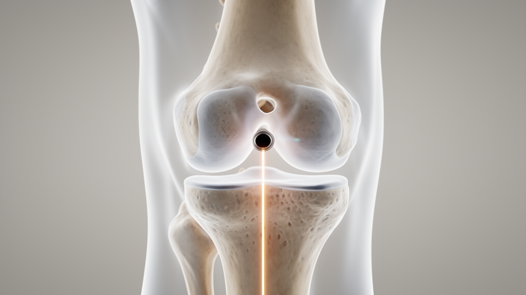
A technique that asks surgeons to do something remarkably simple — glance at the intercondylar notch of the knee before drilling — has passed its first formal accuracy test. In a retrospective cross-sectional study of 108 patients who underwent anterior cruciate ligament reconstruction, researchers found that a notch-referenced check of the tibial guide pin placed the tunnel centre within roughly one millimetre of the patient’s native ligament footprint, measured on three-dimensional imaging. The work, published in Archives of Orthopaedic and Trauma Surgery, offers a potential answer to one of the most stubborn technical problems in knee surgery: getting the tibial tunnel in exactly the right place, left to right, without expensive gadgets.
When surgeons dissect the reasons, non-anatomic tunnel placement consistently emerges as a leading technical contributor, present in up to 77% of revision cases. The graft must sit where the original ligament sat; if it does not, the knee’s natural kinematics are disturbed, the graft experiences abnormal loading, and the risks of impingement and elongation rise. Accurate positioning relative to the native ACL footprint is therefore considered essential to restoring functional stability and protecting long-term outcomes.
The difficulty is not evenly distributed across the anatomy. Positioning the tunnel from front to back — the anteroposterior direction — is comparatively well served by reliable landmarks, including the posterior border of the anterior horn of the lateral meniscus, the posterior margin of the tibial plateau, the posterior cruciate ligament, and any ACL remnant that survives the injury. The mediolateral direction, by contrast, is a blind spot. The references surgeons commonly use, such as the medial tibial eminence or the lateral border of the medial tibial spine, are imperfect, and no distinct, constant bony landmark reliably defines the lateral boundary of the ACL tibial footprint. Mediolateral placement is thus inherently subjective, dependent on arthroscopic visualisation and surgical experience.
Several factors can make matters worse. In chronic ACL-deficient knees, the ligament remnant is often absent, displaced, or scarred, removing a potentially useful guide. Anatomical variability in tibial plateau morphology, including sex- and population-related differences, further undermines landmark-based techniques. The result is that even when anatomic reconstruction is intended, substantial deviations from the native footprint are frequently observed. Adjunctive methods exist — modified portal placement to improve visualisation, computer-assisted navigation, and fluoroscopy-guided tunnel placement — but their routine use is limited by increased cost, operative time, and technical complexity. What the field lacks is a simple, reproducible, widely applicable intraoperative method aimed specifically at the mediolateral axis.
The technique tested in the new study, called Notch-Referenced Mediolateral (NRML) verification, draws on a structure surgeons already see in every case: the intercondylar notch, the consistent anatomical corridor through which the ACL passes. With the tibial guide pin left in situ, the knee is gradually moved from flexion into full extension under arthroscopic visualisation. A reference line, perpendicular to the guide-pin trajectory, is drawn from the lowest point of the medial notch wall across to the lateral notch wall and divided into equal medial, central, and lateral thirds. If the pin crosses the middle third, mediolateral alignment is judged appropriate; if it crosses an outer third, the pin is repositioned before definitive drilling. Only minimal notch debridement for visualisation is permitted — no formal notchplasty — to preserve the notch as a consistent reference.
To validate the approach, the team studied 108 consecutive patients, drawn from roughly 300 screened, who underwent primary all-inside single-bundle ACLR with NRML verification at a single institution between January 2024 and December 2025. All patients received preoperative 3D MRI and immediate postoperative 3D CT under standardised protocols. Two blinded orthopaedic residents independently measured the native tibial footprint centre on MRI and the tibial tunnel centre on CT using a standardised quadrant-grid protocol, repeating the measurements after a week to gauge reliability. The primary outcome was mediolateral deviation, calculated as tunnel position minus footprint position, with negative values indicating medial deviation.
The numbers were striking in their consistency. The anatomical footprint centre sat at 46.1 ± 2.3% of tibial plateau width in the mediolateral direction, while the tunnel centre averaged 44.8 ± 2.0%. Mean deviation was −1.01 ± 1.52 mm (p < 0.001), a small but statistically significant medial bias. Crucially, 91.7% of tunnels (95% CI 84.9–95.6%) fell within ±3 mm of the native footprint centre — a threshold the authors describe as a practical, literature-based benchmark for anatomic placement rather than a strict biological cutoff. MRI–CT agreement was good, with intraclass correlation coefficients of 0.81 to 0.85, and inter- and intraobserver reliability ranged from 0.78 to 0.89, spanning good to excellent.
Accuracy also held steady across patient subgroups. No statistically significant associations were found between mediolateral deviation and sex, injury chronicity, graft diameter, or tibial plateau size. Deviation was numerically smaller in acute injuries (−0.92 ± 1.45 mm) than chronic ones (−1.15 ± 1.65 mm), but the difference did not reach significance. The authors suggest this robustness fits a technique that relies on relative alignment within the notch rather than fixed distances, allowing it to adapt to individual anatomy — though they caution that the subgroup of female patients was small, at just 12 of 108 participants.
How does roughly one millimetre compare with conventional practice? Previous studies using standard landmark-based techniques have reported centre-to-center deviations of approximately 3–4 mm, including 3.50 ± 1.6 mm with independent drilling and 4.40 ± 1.7 mm with transtibial techniques; MRI-based virtual arthroscopy has shown placement errors of about 3 mm. One prior study reported a mean deviation of 6.24 mm, with only 45% of cases achieving at least 50% overlap with the anatomical insertion. Another, using postoperative 3D CT with a footprint reconstructed from bony landmarks within the same modality, found a smaller medial deviation of about 0.67 mm — but the authors note that a cross-modality MRI-to-CT comparison, as used here, is a more demanding test.
One pattern deserves attention: the deviation ran systematically medial rather than scattering evenly around zero. The authors offer a plausible, if untested, explanation — surgeons may instinctively favour a slightly medial guide-pin position as a safety margin, because the anterior horn of the lateral meniscus lies immediately lateral to the ACL tibial footprint and has been flagged as at risk when a tunnel is centred on the footprint. Cadaveric drilling studies support that concern, showing measurable anterior horn injury after reaming, particularly with larger reamers. The authors flag this trade-off as worth tracking as the notch-referenced concept spreads.
The study’s limitations temper enthusiasm appropriately. Its retrospective, single-arm design demonstrates that NRML verification is feasible and radiologically accurate, but only a comparative trial against conventional landmark-based placement can establish whether it offers a genuine advantage. The medial/central/lateral categorisation, though explicitly defined, rests on visual judgment and likely carries a learning curve; all procedures were performed by two experienced surgeons, which aids internal consistency but may limit generalisability. The analysis focused on radiological accuracy and included no clinical outcomes, and imaging-based measurement remains dependent on accurate identification of anatomical landmarks. The authors call for prospective, controlled comparisons and for correlating radiologic accuracy with clinical outcomes to establish clinical relevance.
Even so, the appeal of the technique lies in its simplicity. NRML verification requires no additional equipment, no intraoperative imaging, and no new anatomical landmark — it functions as a dynamic intraoperative check within a standard arthroscopic workflow, performed before definitive drilling. In a single-arm cohort, it placed the tibial tunnel within about one millimetre of the native mediolateral footprint, with 91.7% of tunnels within ±3 mm and high measurement reliability. Whether that radiological precision translates into better knees will take further study, but the notion that the answer to a persistent surgical blind spot might already be visible through the arthroscope — in the notch itself — is a compelling one.
Subject of Research: Validation of a notch-referenced technique for mediolateral tibial tunnel positioning in ACL reconstruction
Article Title: Notch-referenced mediolateral verification of tibial tunnel position in anterior cruciate ligament reconstruction: a 3D imaging validation study
Article References: Nam, V. T., Thang, V. D., Huy, L. D., Phuc, L. H., Anh, L. N., Thinh, L. D., Long, C. H., Huyen, N. N., Trung, N. D., Van Thanh Nam, T., Minh, H. N., & Dung, T. T. (2026). Notch-referenced mediolateral verification of tibial tunnel position in anterior cruciate ligament reconstruction: a 3D imaging validation study. Archives of Orthopaedic and Trauma Surgery, 146(1), Article 368. https://doi.org/10.1007/s00402-026-06442-w
Image Credits: AI Generated
DOI: 10.1007/s00402-026-06442-w
Keywords: ACL reconstruction, tibial tunnel placement, intercondylar notch, 3D MRI, 3D CT, surgical technique, knee surgery, orthopaedics, tunnel accuracy, sports medicine, anatomic reconstruction, Notch-referenced
News Source: Ophelia Keating. (October 30, 2026). Simple notch check guides ACL graft tunnel within a millimetre on 3D imaging. Scienmag.