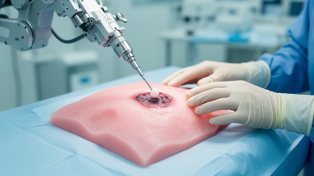
Surgeons often rely on touch to find the edges of a tumor before cutting it out, feeling for tissue that is stiffer than the healthy material around it. That instinct works when the tumor is directly reachable, but it fails during minimally invasive procedures in the head, neck and brain, where fingers cannot follow. A team of engineers at Technische Universität Ilmenau in Germany now reports a proof of concept for a simple, quantitative alternative: a force sensor with a spherical tip that presses into tissue-like material, measures how hard it has to push, and calculates a stiffness value on the spot. In tests on silicone phantoms containing stiff, tumor-like inclusions, the method mapped the shape of hidden inclusions more clearly than experienced surgeons could by manual palpation.
The study, published in BMC Biomedical Engineering, was led by Max Jäger, Katja Uhrhan, Christine Mucha, María Alejandra Guzmán Alfaro and Hartmut Witte. Its central idea is deliberately low-tech compared with many existing approaches. Instead of ultrasound probes, optical systems or robotic grippers packed with electronics, the researchers used a one-axis strain gauge load cell connected to a rigid probe with a 5-millimeter spherical indenter tip. The sensor was calibrated against known weights, achieving a linear fit with a coefficient of determination above 0.999 and a force slope of 0.7658 newtons per volt. Force signals were sampled at 1 kilohertz and processed offline in a Python script.
The physics behind the measurement comes from classical contact mechanics. When a much stiffer sphere indents an elastic half-space, Young’s modulus of the material can be calculated from the normal force, the indentation depth, the radius of the tip and an assumed Poisson’s ratio. The team fixed Poisson’s ratio at 0.5, treating the silicone samples as incompressible during the brief, static indentation, which lasted only about half a second at each point. Both the tip radius and the ratio were treated as constants, so the only live inputs were the measured force and the depth to which the tip sank into the surface.
To create a defined reference, the researchers manufactured silicone phantoms following a published casting process. Four phantoms were used: one stiff, one soft, and two with stiff inclusions embedded in soft surrounding material, one cylindrical and one star-shaped. The soft material was Ecoflex 00-20 silicone rubber and the stiff material was a transparent Shore A-30 silicone. The stiff material’s modulus was estimated from Shore hardness equations at roughly 1.14 to 1.15 megapascals, while the soft material’s modulus was taken as about 51 kilopascals based on four literature studies, with a standard deviation of 9.54 kilopascals across those published means. The phantoms were 85 millimeters in diameter and roughly 15 millimeters thick, and the team kept deformations below 10 percent to stay within the linear-elastic regime where the calculation is valid.
Validation began with repeated measurements at a single point on each homogeneous phantom, ten measurements per material at an indentation depth of 0.5 millimeters. The results showed strong repeatability: the standard deviation of the estimated modulus was 3.4 percent of the mean for the soft material and 6.7 percent for the stiff one, both below 7 percent. Accuracy told a more nuanced story. On the soft phantom, the estimated modulus of about 64 kilopascals slightly overshot the literature maximum of 60 kilopascals, a relative error of 6.7 percent. On the stiff phantom, the mean estimate fell far below the reference, with a relative error of 44.9 percent. The authors attribute this mainly to errors in indentation depth: the stiffer the material, the more an incorrect depth distorts the calculated modulus, because measured force in stiff materials depends much more strongly on depth than it does in soft ones.
The contact point between tip and surface was identified visually by the investigator, which the team identifies as the likely source of those depth errors. Interestingly, that visual judgment proved inaccurate but reproducible, which is why the standard deviations stayed low even as absolute accuracy suffered on the stiff material. The researchers calculated that the indentation depth uncertainty must stay below 0.037 millimeters to cleanly separate materials whose moduli differ by a factor of two, such as the 50 and 100 kilopascals they assumed for soft tissue and a stiff inclusion buried under 7 millimeters of soft material. That threshold depends on the stiffness contrast being targeted and would need recalculating for other use cases.
The real test was tissue discrimination. The team scanned a line of 15 points across the diameter of a phantom with a cylindrical stiff inclusion, once with the inclusion about 1 millimeter below the surface and once with about 7 millimeters of soft material above it, achieved simply by flipping the phantom over. In both configurations, a Mann-Whitney-Wilcoxon test found a statistically significant difference in Young’s modulus between points above the inclusion and points above soft material, at a significance level of 5 percent. Even buried under 7 millimeters of soft silicone, the stiff inclusion left a detectable fingerprint in the stiffness profile, though the transition was softened, which the authors liken to an image blurred by frosted glass.
The most striking comparison came from surface scans versus human hands. Two scans were performed on a phantom with a star-shaped inclusion: one covering roughly half the surface on a 2-millimeter grid, and a finer one covering a single star tip on a 1-millimeter grid. Both scans captured the overall shape of the inclusion, with the contour far clearer in the higher-resolution scan. After omitting some erroneous measurement points caused by inaccurate contact detection, a Mann-Whitney-Wilcoxon test comparing 180 points inside the inclusion outline with 270 points outside yielded a p-value below 10 to the minus 6, a Wilcoxon statistic of 584 and a Cohen’s d of 3.311, indicating a very large effect. Meanwhile, six experienced surgeons from ENT and neurosurgery departments palpated three phantoms with differently shaped inclusions through a cardboard box and drew what they felt. The circular inclusion was detected correctly, but both star-shaped inclusions proved difficult. The authors conclude it seems safe to assume their method provides better inclusion shape detection than manual palpation, while noting the comparison is a first impression from a small exploratory study.
The experiment with surgeons was approved by the Ethics Committee of Technische Universität Ilmenau and conducted in accordance with the Declaration of Helsinki, with written informed consent from all participants. The authors are careful to frame the work as a technical validation rather than a clinical result. The setup is a laboratory bench system with manual positioning on three linear stages, stepper-motor-driven table movement and depth read from a fine-thread screw. It is suitable for laboratory experimentation and ex-vivo tissue analysis, but not yet for use alongside resection in a living patient. For that, the probe would need to be miniaturized, easier to handle, and compatible with handheld use or attachment to a surgical robot, with tracking that does not hinder the surgeon’s movements.
Several engineering hurdles remain before the technique could reach the operating room. In minimally invasive ENT and neurosurgery, the indenter often cannot approach the tissue perpendicularly, so the authors argue that torque and lateral force sensing, perhaps with a 6-axis force/torque sensor, will be needed. Automated, feedback-controlled indentation depth detection is another priority, both to speed up measurements and to keep deformation within the 10 percent limit that protects biological tissue from damage and preserves linear-elastic behavior. Possible solutions from the literature include capturing the surface topography before indentation, using a transparent tip to infer depth from the contact area, and measuring tool deflection while controlling indentation force. On sterilization, the design offers an advantage: only the removable indenter tip touches tissue, so the force sensor can be covered during surgery and just the tip sterilized. The team plans measurements on ex-vivo biological tissue samples, including tumor tissue, to test whether the phantom performance transfers to clinical application, forming the basis for future in-vivo adaptation.
Subject of Research: An indentation-based sensing method for determining mechanical tissue properties to locate tumor boundaries during surgery
Article Title: Proof of concept of a static approach to determine mechanical tissue properties during tumor surgery
Article References: Jäger, M., Uhrhan, K., Mucha, C., Guzmán Alfaro, M. A., & Witte, H. (2025). Proof of concept of a static approach to determine mechanical tissue properties during tumor surgery. BMC Biomedical Engineering, 7(1), Article 15. https://doi.org/10.1186/s42490-025-00100-9
Image Credits: AI Generated
DOI: 10.1186/s42490-025-00100-9
Keywords: tumor surgery, tissue stiffness, Young's modulus, instrumented palpation, silicone phantoms, tumor boundaries, minimally invasive surgery, force sensing, contact mechanics, ENT surgery, neurosurgery, biomedical engineering
News Source: Denise Maddox. (October 30, 2026). Robot-assisted indentation outperforms surgeon palpation in tumor boundary test. Scienmag.