


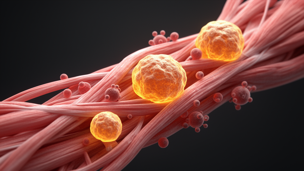
Skeletal muscle has an extraordinary ability to rebuild itself after injury, and a new review argues that one of the most surprising players in that process is cellular senescence—the stress-induced state in which cells permanently stop dividing. Writing in the journal Cell Regeneration, researchers Xingyuan Liu and Huating Wang survey years of evidence showing that senescent cells can act as powerful allies of muscle regeneration when they appear briefly, yet become obstacles to repair when they linger. The review, published on 7 May 2026, synthesizes a field that has produced genuinely contradictory results, and it lays out why resolving those contradictions matters for developing therapies against age-related muscle decline and degenerative disease.
The authors frame senescence as a paradoxical regulator rather than a simple villain. In their abstract, they describe accumulating evidence that transient senescence—particularly in fibro-adipogenic progenitors, macrophages, and other niche cells during acute muscle injury—plays a beneficial role in supporting regeneration. These senescent cells promote cellular plasticity, enhance myoblast differentiation, facilitate phagocytic clearance of debris, and modulate inflammation through the timely secretion of senescence-associated secretory phenotype factors, the chemical cocktail known as the SASP. But conflicting findings suggest the opposite: when senescence persists in aged or dystrophic muscle, senescent cells impair regeneration by establishing a sustained pro-inflammatory and pro-fibrotic niche.
To understand why senescence matters here, it helps to know how muscle repair normally unfolds. Skeletal muscle makes up roughly 30 to 40 percent of human body mass, and its regeneration depends primarily on muscle stem cells, or MuSCs, which sit quiescently between the basal lamina and the sarcolemma, held in check by PAX7-dependent transcription and cell-cycle inhibitors. When injury strikes, damaged myofibers undergo necrosis and trigger an inflammatory response. Neutrophils infiltrate within hours, clearing debris and recruiting macrophages. Pro-inflammatory M1 macrophages then phagocytose necrotic material and secrete factors that activate MuSCs and drive myoblast proliferation. Later, a shift toward anti-inflammatory M2 macrophage polarization—partly orchestrated by regulatory T cells secreting IL-10, IL-13, and IL-4—allows inflammation to resolve and myoblasts to differentiate and fuse into new myofibers.
MuSCs do not act alone. Fibro-adipogenic progenitors, marked by PDGFRα expression, rapidly expand after acute injury and secrete growth factors such as WISP1 and IGF-1 that directly activate MuSCs. They also help phagocytose necrotic debris and produce extracellular matrix components that structurally support repair. Endothelial cells promote angiogenesis and preserve MuSC quiescence by secreting the Notch ligand Dll4, while MuSCs reciprocate with VEGF-A to stimulate vascular growth. Pericytes stabilize nascent vessels and secrete IGF-1 to promote myoblast differentiation, and neural cells help re-establish neuromuscular junctions. In chronic injury states such as Duchenne muscular dystrophy, however, this coordinated system breaks down: reparative inflammation shifts into a chronic, maladaptive state that damages healthy fibers, impairs MuSC function, and promotes excessive fibrosis.
Cellular senescence itself is a state of stable, essentially irreversible cell-cycle arrest induced by stressors including telomere dysfunction, DNA damage, oncogene activation, oxidative stress, and mitochondrial impairment. First described by Hayflick and Moorhead in 1961 as replicative senescence in human fibroblasts, the phenomenon is enforced through the p53/p21 and p16INK4a/pRB tumor suppressor pathways. Senescent cells acquire distinctive features: hypertrophy and flattening, elevated senescence-associated β-galactosidase activity, loss of nuclear lamin B1, accumulation of lipofuscin granules, persistent DNA damage foci, and the sustained release of SASP factors such as IL-6, IL-8, CCL2, TGF-β, and matrix metalloproteinases. The SASP is regulated by signaling nodes including NF-κB, p38MAPK, mTOR, C/EBPβ, and AP-1 transcription factors.
Senescence is not always harmful. The review highlights its programmed, beneficial roles in embryonic development, where p21-dependent senescent cells secrete FGF ligands to guide tissue patterning and are then cleared by macrophages. In skin wound healing, transient senescence in fibroblasts and endothelial cells promotes myofibroblast differentiation and angiogenesis through SASP-mediated secretion of PDGF-AA and VEGF; selectively eliminating p16INK4a-positive cells in p16-3MR mice delays wound closure, an effect rescued by exogenous PDGF-AA. In the liver, CCN1-induced senescent stellate cells secrete matrix metalloproteinases that promote remodeling and inhibit fibrosis, and in the heart, transient senescence in fibroblasts supports regeneration by enhancing cardiomyocyte proliferation and reducing fibrosis. These regenerative effects depend critically on the transient nature of the senescent state; prolonged persistence shifts the SASP toward a pro-inflammatory, pro-fibrotic profile that impairs repair.
In skeletal muscle, the earliest evidence for a pro-regenerative role came in 2015, when Le Roux and colleagues first demonstrated senescence induction in tibialis anterior muscle under both acute cardiotoxin injury in young mice and chronic injury in mdx mice, a Duchenne model. More than half of the senescent cells expressed the endothelial marker Flk-1, and the cells were absent in healthy muscle but accumulated transiently during early regeneration. In 2017, Chiche and colleagues reported that injury-induced senescent cells, acting through SASP factors—particularly IL-6—promote a partial reprogramming state in Pax7-positive MuSCs that increases cellular plasticity and improves regenerative capacity, without progressing to full pluripotency or teratoma formation. Eliminating senescent cells with the senolytic drug ABT-263 or in p16-3MR mice reduced both senescent cell burden and plasticity. Then, in 2020, Saito and colleagues identified senescent FAPs as a predominant population four days after acute injury; these cells promoted macrophage-mediated phagocytic clearance, enhanced myoblast differentiation in co-culture, and, when transplanted into injured muscle in vivo, enhanced regeneration and reduced fibrosis.
Single-cell and spatial mapping technologies have since sharpened the picture. Young and colleagues showed that in acutely injured muscle, senescence and SASP gene activity localize precisely to zones of active repair and inflammation, with senescent cells appearing around three days post injury and declining to undetectable levels by 21 days. Eliminating these cells with ABT-263 impaired repair, reducing myofiber cross-sectional area and Pax7-positive MuSC abundance. In chronically injured mdx mice, by contrast, senescent cells accumulated persistently. Walter and colleagues developed a senescence scoring method revealing a transitory senescent MuSC population abundant at 3.5 days post injury across all ages but increased in older mice and associated with impaired regenerative capacity. A unified senescence score applied to human muscle identified senescent MuSCs, endothelial cells, smooth muscle cells, and FAPs in young tissue, significantly increased in aged muscle.
The contradictory evidence is equally striking. Dungan and colleagues found that senescent cells—predominantly CD11b-positive macrophages—accumulate after injury in both young and old mice, but in aged mice they persist and express elevated senescence and SASP genes, correlating with impaired regeneration. Clearing them with the senolytic combination dasatinib and quercetin enhanced regeneration in old mice, producing larger myofiber cross-sectional area, increased MuSC abundance, and improved physical function. Yet the same treatment impaired regeneration in young mice, reducing fiber size and MuSC activation. More recently, Moiseeva and colleagues argued that even transiently emerging senescent cells in acutely injured young muscle are harmful: using live-cell staining and single-cell sequencing, they showed that senescent FAPs, myeloid cells, and MuSCs create an aged-like, inflamed niche through pro-fibrotic and pro-inflammatory SASPs that disrupts MuSC proliferation, and that clearing these cells enhanced regenerative outcomes, reduced fibrosis and inflammation, and restored muscle function in both acute and chronic injury models.
The review’s authors argue that these conflicts likely stem from unresolved gaps rather than irreconcilable data. Senescent cells in regenerating muscle are heterogeneous, and global clearance strategies such as ABT-263, dasatinib plus quercetin, and the p16-3MR model—which itself has been shown to clear senescent cells inefficiently—eliminate mixed populations whose individual contributions are conflated. Distinguishing transient senescence from the persistent senescence of aged or dystrophic muscle, tracking the fate of senescent cells over time, and identifying the upstream signals that initiate senescence in the regenerating niche are, the authors contend, the urgent next steps. Only then, they conclude, can targeted senotherapies be designed to eliminate deleterious subsets while preserving the pro-regenerative ones—potentially enhancing muscle regeneration in aging and degenerative disease.
Subject of Research: The paradoxical roles of cellular senescence in skeletal muscle regeneration
Article Title: Cellular senescence in skeletal muscle regeneration
Article References: Liu, X., & Wang, H. (2026). Cellular senescence in skeletal muscle regeneration. Cell Regeneration, 15(1), Article 15. https://doi.org/10.1186/s13619-026-00287-9
Image Credits: AI Generated
DOI: 10.1186/s13619-026-00287-9
Keywords: cellular senescence, skeletal muscle, muscle regeneration, muscle stem cells, SASP, fibro-adipogenic progenitors, senolytics, aging, Duchenne muscular dystrophy, macrophages, single-cell RNA sequencing, Cell Regeneration
News Source: Beatrice Stafford. (October 30, 2026). Senescent Cells Play Both Hero and Villain in Muscle Repair, Review Finds. Scienmag.



