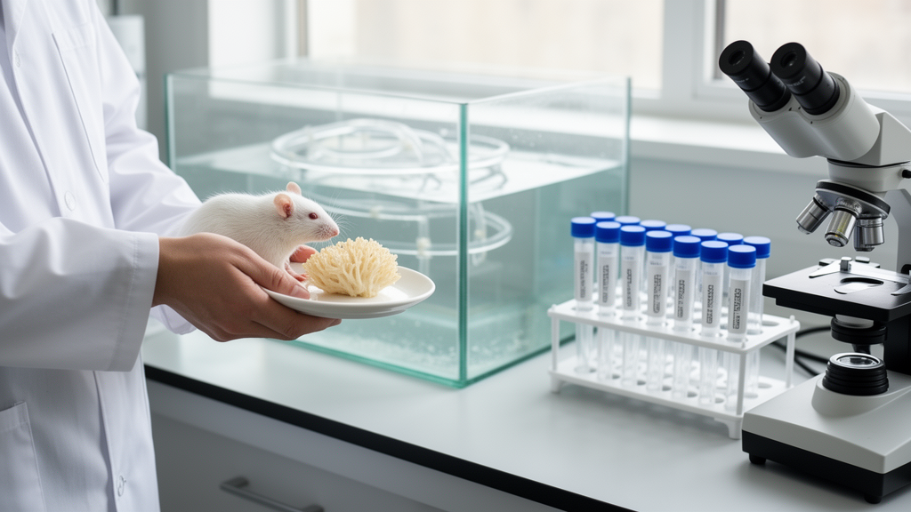
A polysaccharide extracted from the lion’s mane mushroom, Hericium erinaceus, has been found to partially reverse the metabolic, intestinal, and memory-related damage caused by a high-fat, high-sucrose diet in mice, according to a new study published in Frontiers in Nutrition. The research, led by Wei Deng of the People’s Hospital of Yuechi County and colleagues at Southwest Medical University in China, suggests that this mushroom-derived compound may act on the gut-brain axis, the communication network linking the intestinal tract to the central nervous system, though the findings so far come exclusively from animal experiments.
The team fed male C57BL/6J mice a high-fat, high-sucrose diet for 42 days, with one group of diet-fed animals additionally receiving daily oral gavage of the polysaccharide, known as HEP, at a dose of 300 milligrams per kilogram of body weight. A normal-chow control group and a metformin-treated positive control group were included for comparison. Over the course of the study, the researchers ran glucose tolerance and insulin tolerance tests at weeks four and five, measured serum lipids, assessed spatial working memory with a Y-maze test in the final week, sequenced gut bacterial communities from colon contents using 16S rRNA gene sequencing, and examined colon and hippocampal tissues with histological staining, ELISA assays, and Western blotting.
The dietary intervention produced the expected damage. Mice on the high-fat, high-sucrose diet showed elevated blood glucose during the glucose tolerance test, a blunted hypoglycemic response to insulin indicating insulin resistance, and dyslipidemia marked by raised triglycerides, total cholesterol, and LDL cholesterol alongside a downward trend in HDL. In the Y-maze test, these animals displayed a significantly lower spontaneous alternation rate, a standard measure of spatial working memory, and their movement traces suggested reduced exploration and activity compared with the normal-chow controls.
HEP treatment partially rescued these deficits. The treated mice showed attenuated glucose elevations during the tolerance test, with area-under-the-curve values comparable to the control group, a stronger glucose-lowering response to insulin, and reduced serum triglycerides, cholesterol, and LDL with partial restoration of HDL. On the behavioral task, the polysaccharide significantly increased the spontaneous alternation rate relative to the untreated diet-fed mice, with a statistical significance level of p < 0.01, and the metformin positive control produced a comparable reversal, confirming that the model responded to cognitive-improving interventions.
Sequencing of the gut microbiota revealed that the diet reshaped bacterial community structure without significantly changing overall species richness. Alpha-diversity indices such as ACE, Chao1, and Shannon showed downward trends in the diet-fed group that did not reach statistical significance, and HEP supplementation produced only non-significant upward trends. Beta-diversity analysis, however, showed clear separation among the three groups by both principal coordinate and non-metric multidimensional scaling ordination, with a stress value of 0.038 confirming the reliability of the ordination model. The HEP-treated mice also harbored 306 unique amplicon sequence variants compared with 109 in the untreated diet group, suggesting greater ecological complexity after intervention.
Differential abundance analysis identified distinct signature taxa in each group. Lactobacillus and members of the Lactobacillaceae family characterized the normal-chow controls, while Candidatus Stoquefichus was enriched in the diet-only mice. In the HEP-treated group, the discriminatory taxa were concentrated within the phylum Actinobacteria and included Corynebacterium, Psychrobacter, Jeotgalicoccus, Acinetobacter, and Glutamicibacter. Notably, the authors acknowledge that this pattern differs from earlier HEP studies that reported enrichment of genera such as Lactobacillus and Bifidobacterium, a divergence they attribute to differences in disease context, polysaccharide preparation, dosing regimen, and host species, and they emphasize that in an already imbalanced intestinal environment the primary effect of HEP appeared to be holistic remodeling of community architecture rather than enrichment of specific probiotic genera.
In the colon, the diet caused visible damage: H&E staining revealed crypt shortening, disorganized epithelial structure, and increased inflammatory cell infiltration, while PAS staining showed significantly fewer goblet cells and weaker staining, indicating impaired mucus production. Immunohistochemistry showed that the tight junction proteins Occludin and ZO-1, which seal the gaps between epithelial cells, were sparse and discontinuous in the diet-fed mice. HEP intervention improved mucosal architecture, partially restored goblet cell numbers, and increased both the intensity and continuity of Occludin and ZO-1 staining, although goblet cell counts did not fully return to the levels seen in normal-chow controls.
The hippocampus, a brain region central to learning and memory, showed parallel changes. ELISA measurements revealed that diet-fed mice had significantly decreased levels of the neurotransmitter serotonin (5-HT) and the neurotrophic factor BDNF, along with elevated levels of the pro-inflammatory cytokines TNF-α and IL-6. HEP treatment significantly increased 5-HT and BDNF while reducing TNF-α and IL-6. Western blot analysis added a signaling-level picture: the ratio of phosphorylated to total NF-κB p65, a marker of inflammatory pathway activation, was significantly elevated in the diet group and significantly reduced by HEP, while the phosphorylated-to-total CREB ratio, linked to neuroplasticity and BDNF expression, was significantly decreased by the diet and restored by the intervention. In both cases the HEP-treated group did not differ significantly from the metformin group.
The authors propose a mechanistic chain connecting these observations. A compromised intestinal barrier allows bacterial products such as lipopolysaccharide to translocate into the circulation, triggering low-grade systemic inflammation that can reach the brain through the blood-brain barrier or vagal pathways and drive neuroinflammation, which in turn suppresses hippocampal BDNF and 5-HT systems and impairs synaptic plasticity and memory. They cite prior work showing that fecal lysate from high-fat-diet mice can directly suppress BDNF expression and CREB phosphorylation, an effect partially blocked by an anti-LPS antibody, and note that most serotonin is synthesized in intestinal enterochromaffin cells under microbial influence. In their own data, the restoration of barrier-related indices ran in close parallel with the reduction of hippocampal inflammatory markers.
Important caveats accompany these findings. The study was conducted entirely in mice, so translational relevance to humans remains untested, and the authors explicitly state that they did not perform causal validation experiments such as fecal microbiota transplantation or antibiotic depletion in their own animals, nor did they use pathway inhibitors to block downstream inflammatory signaling. The absence of immunofluorescence staining for microglial and astrocyte activation markers also limits the mechanistic depth of the work. The authors frame the results as an association-based foundation for future studies targeting the microbiota-gut-brain axis in metabolic disorder-related cognitive impairment, with planned follow-up work including short-chain fatty acid quantification and pharmacological blockade of TLR4 signaling. Until such causal and human studies are completed, the mushroom polysaccharide should be regarded as a promising but unproven candidate rather than an established therapy.
Subject of Research: Effects of Hericium erinaceus polysaccharide on gut microbiota, intestinal barrier integrity, and spatial working memory in high-fat high-sucrose diet-fed mice
Article Title: Hericium erinaceus polysaccharide ameliorates spatial working memory impairment and intestinal barrier dysfunction in high-fat high-sucrose diet-fed mic
Article References: Deng, W., Han, Y., Yang, H., Mao, S., Huang, Q., Tang, Q., Zhang, Y., Tang, S., Li, R., Cui, X., Huang, Z., Wang, X., Wang, X., Wang, J., He, B., & Wang, X. (2026). Hericium erinaceus polysaccharide ameliorates spatial working memory impairment and intestinal barrier dysfunction in high-fat high-sucrose diet-fed mic. Frontiers in Nutrition, 13, 1898939. https://doi.org/10.3389/fnut.2026.1898939
Image Credits: AI Generated
DOI: 10.3389/fnut.2026.1898939
Keywords: Hericium erinaceus, polysaccharides, gut-brain axis, gut microbiota, intestinal barrier, high-fat high-sucrose diet, spatial working memory, hippocampus, neuroinflammation, BDNF, mouse study, Frontiers in Nutrition
News Source: Daisy Hatcher. (October 27, 2026). Research report: Hericium erinaceus polysaccharide ameliorates spatial working memory impairment and intestinal barrier dysfunction in high-fat high-sucrose diet-fed mic. Scienmag.