A 23-year-old woman with insulin resistance and severe obesity arrived at an emergency department in Beirut with severe upper abdominal pain, nausea and vomiting that had begun a day earlier. Blood tests told a striking story: her serum triglycerides measured 7,533 mg/dL, far above any safe threshold, and her lipase was elevated at 292 U/L. A computed tomography scan of the abdomen showed mild acute interstitial pancreatitis with inflammatory changes around the pancreas, but no gallstones, no pancreatic necrosis and no fluid collections. She was diagnosed with hypertriglyceridemia-induced acute pancreatitis and admitted to the intensive care unit with an initial BISAP severity score of 1.
What happened next is described in a case report published in Frontiers in Medicine on 28 October 2026 by physicians at the American University of Beirut. Within hours, the young woman’s condition unraveled. Ten hours after admission she developed sinus tachycardia reaching 160 beats per minute and tachypnea up to 35 breaths per minute, followed by a high-grade fever twenty hours after admission. Repeat laboratory testing showed hemoconcentration, acute kidney injury with creatinine rising to 1.44 mg/dL, persistent metabolic acidosis and severe hypocalcemia with a calcium level of 6 mg/dL. She received additional intravenous fluids, calcium gluconate and empiric piperacillin-tazobactam while blood cultures were drawn, even though her chest X-ray and urinalysis showed no infection source.
Thirty-six hours after admission came the first neurological alarm. She became agitated, disoriented and delusional, at one point insisting she was pregnant. She then developed seizure-like activity with upward and rightward eye deviation, tonic posturing and unresponsiveness. Her serum calcium around the seizure had fallen to 5.6 mg/dL, and clinicians treated the episode as a provoked seizure, giving intravenous diazepam and loading doses of levetiracetam. Because she remained unresponsive for fifteen minutes and developed respiratory failure, she was intubated. Hypotension followed, requiring vasopressor support. Neuroimaging was considered at that moment but deferred because she was too hemodynamically unstable to transport, needing two pressors to maintain her blood pressure.
Her metabolic picture continued to deteriorate. Post-intubation blood gases showed worsening metabolic acidosis with a bicarbonate of 8 mmol/L and an arterial pH of 7.16, and her creatinine climbed to 3 mg/dL. Triglycerides, notably, were falling fast, dropping to 1,419 mg/dL and then to 617 mg/dL under insulin therapy. Continuous renal replacement therapy was started for progressive kidney injury, oliguria and refractory acidosis, and a normothermia protocol was initiated for a persistent temperature of 41 degrees Celsius. Neurologically, however, she slipped into a severely depressed mental state with no motor response to painful stimuli, even as her brainstem reflexes were preserved.
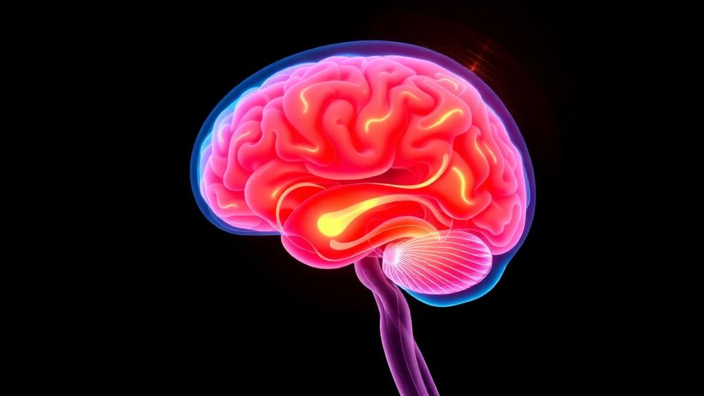
The next day brought the catastrophic finding. Magnetic resonance imaging showed diffuse cerebral and cerebellar edema with extensive restricted diffusion involving the white matter of both the cerebrum and cerebellum. Magnetic resonance angiography demonstrated absent flow-related enhancement within the cervical and intracranial arterial circulation, and electroencephalography showed a severely suppressed background with no identifiable cerebral activity above noise at a sensitivity of 70 µV/cm. Hyperosmolar therapy with mannitol and hypertonic saline was begun, but it was withheld shortly afterward as futile given the extent of the injury.
The clinical team attributed the deterioration to pancreatic encephalopathy, a rare and poorly understood neurological complication of severe acute pancreatitis first described in 1941. The diagnosis, however, is one of exclusion, and the authors systematically ruled out competing explanations. Severe hypocalcemia may have provoked the seizure, but it was promptly corrected and could not plausibly explain diffuse cerebral edema and herniation. Kidney injury, metabolic acidosis, sepsis, high fever and a post-ictal state were considered, but none accounted for the catastrophic course. Infectious meningoencephalitis was judged less likely given the absence of preceding infectious symptoms and imaging showing diffuse global injury rather than a focal process. The chart review found no sustained oxygen desaturation or prolonged hypotension that would suggest hypoxic injury.
The temporal pattern of the deterioration fits what is known about the condition. Pancreatic encephalopathy is described as the onset of neuropsychiatric symptoms, including altered mental status, psychomotor agitation, hallucinations, seizures, focal deficits and coma, usually within the first two weeks of pancreatitis. Early forms occur as part of multiorgan failure during the inflammatory phase, while later forms are linked to thiamine deficiency from prolonged fasting and vomiting. Reported incidence rates among patients with acute pancreatitis range wildly, from 0.04 percent to as high as 11 percent depending on the population studied, a spread that reflects the absence of any accepted diagnostic criteria. Mortality is high, with reported rates between 43 and 70 percent, driven mainly by multiorgan failure, although among survivors neurological recovery is often favorable.
The proposed mechanisms are complex and remain incompletely understood. Researchers have suggested that activated pancreatic enzymes such as trypsin and phospholipase A2 degrade membrane phospholipids and disrupt the blood-brain barrier, producing cytotoxic and vasogenic cerebral edema. The systemic inflammatory response of severe pancreatitis releases proinflammatory cytokines, including tumor necrosis factor-alpha, interleukin-1 beta, interleukin-6 and interleukin-10, which contribute to endothelial dysfunction, increased vascular permeability and neurological injury. Oxidative stress, hypoxemia, metabolic derangements and microvascular injury may promote white matter demyelination and diffuse brain damage. More recent work supports a bidirectional pancreas-brain axis in which blood-brain barrier disruption primes the brain for neuroinflammation and a self-amplifying cascade of neuro-glial-immune interactions.
The authors believe this is the first reported case of brain herniation secondary to pancreatic encephalopathy. A recent national cohort reported cerebral edema in only 0.04 percent of patients with acute pancreatitis, and only three published case reports, including one by Gill and colleagues in 2017 involving hypertriglyceridemia-induced disease, have clearly documented pancreatic encephalopathy with radiologically confirmed diffuse cerebral edema. The contrast in outcomes is instructive: in Gill’s case, reduction of triglycerides through insulin and plasmapheresis was associated with radiographic improvement and complete neurological recovery, whereas in the present patient neurological decline continued even as triglycerides fell from 7,533 mg/dL to below 1,500 mg/dL within 48 hours and later normalized. This suggests that once extensive blood-brain barrier disruption and cerebral edema develop, injury may progress independently of serum triglyceride levels. The authors also note that obesity, particularly a body mass index above 40 kg/m2, and insulin resistance predispose patients to severe pancreatitis and multiorgan failure, and their patient’s BMI was 40.9 kg/m2, though obesity has not been established as an independent risk factor for the encephalopathy itself.
Three months after presentation, the patient remained intubated in a coma with no meaningful neurological recovery. The authors are candid about the limits of their conclusions: because this is a single case diagnosed largely by exclusion, a definite causal relationship between the pancreatitis and the neurological deterioration could not be established, the contributions of hypocalcemia, seizure, sepsis and metabolic derangement could not be fully excluded, and no incidence, generalizable risk factors or treatment standards can be drawn from one patient. Management of pancreatic encephalopathy remains largely supportive, centered on treating the underlying pancreatitis, correcting metabolic abnormalities and supporting failing organs, while cerebral edema management is extrapolated from neurocritical care guidelines for other conditions. Their central message is practical: new neuropsychiatric manifestations in a patient with severe acute pancreatitis should prompt urgent neurological assessment and early neuroimaging whenever clinically feasible, because delayed recognition of evolving cerebral edema may close the window in which treatment could matter.
Subject of Research: A case report of fulminant pancreatic encephalopathy with malignant cerebral edema and herniation in severe hypertriglyceridemia-induced acute pancreatitis.
Article Title: Fulminant pancreatic encephalopathy with brain edema and herniation in severe hypertriglyceridemia-induced pancreatitis: a case report
Article References: Aboudargham, N., El Akach, N., Hamieh, N., Rammal, A., Barada, K., & Bouakl, I. (2026). Fulminant pancreatic encephalopathy with brain edema and herniation in severe hypertriglyceridemia-induced pancreatitis: a case report. Frontiers in Medicine, 13, Article 1968843. https://doi.org/10.3389/fmed.2026.1968843
Image Credits: AI Generated
DOI: 10.3389/fmed.2026.1968843
Keywords: pancreatic encephalopathy, acute pancreatitis, hypertriglyceridemia, cerebral edema, brain herniation, case report, severe acute pancreatitis, blood-brain barrier, multiorgan failure, neurocritical care, critical care, neuroinflammation
News Source: Ophelia Keating. (October 29, 2026). Research report: Fulminant pancreatic encephalopathy with brain edema and herniation in severe hypertriglyceridemia-induced pancreatitis: a case report. Scienmag.