A 61-year-old man with chronic hepatitis B virus infection presented with upper gastrointestinal bleeding, and the subsequent diagnostic workup led clinicians to an exceptionally rare conclusion: extracavitary primary effusion lymphoma occurring in an HIV-negative patient. Laboratory tests revealed normocytic, normochromic anaemia with a haemoglobin of 7.1 g/dL. Serology showed nonreactive HBsAg with reactive anti-HBc and anti-HBs antibodies, consistent with the patient’s known hepatitis B history, while tests for hepatitis C virus, hepatitis A virus IgM, and HIV-1/2 were all nonreactive. The absence of HIV infection is what makes this case noteworthy, because primary effusion lymphoma is described as an exceptionally rare subtype of non-Hodgkin lymphoma in patients without HIV.
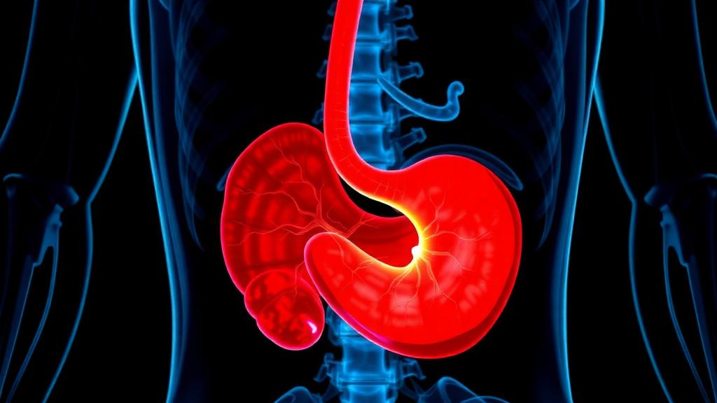
Upper gastrointestinal endoscopy exposed an ulcero-infiltrative lesion in the stomach that involved the entire lesser curvature and the anterior wall of the gastric body, extending to the incisura angularis. The lesion showed extensive necrosis and multiple visible vessels, although without active bleeding at the time of examination. A biopsy was obtained from the affected tissue, setting in motion the histopathological and immunophenotypic analyses that would ultimately define the tumour’s unusual identity.
Under the microscope, the gastric mucosa was densely and diffusely infiltrated by an ulcerated atypical lymphoid population that permeated the gastric glands. The neoplastic cells displayed immunoblastic and plasmablastic morphology, characterized by enlarged irregular nuclei, conspicuous nucleoli, and abundant basophilic cytoplasm. Recognizing this morphology is described as essential for diagnosis, together with demonstration of human herpesvirus 8 and terminal B-cell differentiation markers.
Immunohistochemistry added further complexity. The tumour cells were positive for CD43 and CD45, though with partial loss of the latter, one of the features that can make diagnosis challenging in this lymphoma subtype. Strikingly, the pan-B-cell markers CD20, PAX5, and CD79a were all negative, meaning the tumour lacked the markers that normally identify B-cell lineage. Diffuse CD30 positivity was observed, and the cells showed weak and focal CD3 expression despite being negative for the other T-cell markers CD2, CD5, CD7, CD4, and CD8. The neoplasm expressed MUM1 and EMA but lacked CD138, and stains for ALK, granzyme B, TIA1, CD10, and BCL6 were negative.
The decisive evidence came from viral studies. Epstein–Barr virus was detected by EBER in situ hybridization, and human herpesvirus 8 was demonstrated by LANA-1 immunohistochemistry. The Ki-67 proliferation index exceeded 95 percent, reflecting a very high rate of cellular proliferation. Taken together with the morphology and immunophenotype, these findings established the diagnosis of extracavitary primary effusion lymphoma in this HIV-negative patient.
Primary effusion lymphoma is an exceptionally rare subtype of non-Hodgkin lymphoma in patients without HIV infection. According to the report, the disease may occur in other settings of immune dysfunction, including aging and chronic infections, particularly when it presents as solid extracavitary masses. The patient’s chronic hepatitis B infection is a chronic infection documented in the case, although the report does not establish a causal relationship between the viral hepatitis and the lymphoma.
Diagnosis of this entity is challenging for several reasons documented in the case. Partial loss of CD45 expression, the absence of pan-B-cell markers, and occasional aberrant CD3 expression can all complicate the diagnostic interpretation. The authors emphasize that recognition of immunoblastic and plasmablastic morphology, together with demonstration of human herpesvirus 8 and terminal B-cell differentiation markers, is essential for reaching the correct diagnosis. In the present case, the loss of CD138 expression constituted an additional pitfall, which the authors state highlights the importance of employing a broad immunohistochemical panel when evaluating challenging haematolymphoid neoplasms.
Both viruses detected in this tumour are documented features of the case: Epstein–Barr virus was identified by EBER in situ hybridization, and human herpesvirus 8 was demonstrated by LANA-1 immunohistochemistry. The near-total proliferative activity, with a Ki-67 index above 95 percent, was recorded alongside these viral findings. The combination of morphological, immunophenotypic, and viral evidence was what allowed the diagnosis of extracavitary primary effusion lymphoma to be established in this patient.
The case documents how an HIV-negative patient with an ulcerated gastric mass was found to harbour a virus-associated lymphoma requiring an extensive panel of immunohistochemical stains and viral detection methods for correct classification. The authors note that loss of CD138, as observed in their case, represents an additional pitfall in the diagnostic evaluation of such neoplasms.
Subject of Research: Extracavitary primary effusion lymphoma diagnosed in an HIV-negative patient
Article Title: Extracavitary Primary Effusion Lymphoma in an HIV‐Negative Patient With Aberrant Immunophenotypic Features
Article References: Centoducatte, G. L., Deininger, M. D. G., Lorand‐Metze, I., & Assis‐Mendonça, G. R. (2026). Extracavitary Primary Effusion Lymphoma in an HIV‐Negative Patient With Aberrant Immunophenotypic Features. eJHaem, 7(5), Article e70443. https://doi.org/10.1002/jha2.70443
Image Credits: AI Generated
DOI: 10.1002/jha2.70443
Keywords: primary effusion lymphoma, HHV8, Epstein-Barr virus, HIV-negative, non-Hodgkin lymphoma, immunohistochemistry, plasmablastic morphology, gastric lymphoma, CD30, diagnostic pathology, extracavitary, case report
News Source: Nathaniel Bowman. (October 24, 2026). Rare HIV-Negative Case of Extracavitary Primary Effusion Lymphoma Diagnosed in the Stomach. Scienmag.