Newcastle disease virus, an avian paramyxovirus that infects more than 250 animal species ranging from poultry to mammals, builds conspicuous cytoplasmic inclusion bodies during infection. These membraneless structures have long served as a histopathological hallmark of paramyxovirus disease, yet the molecular rules governing their assembly have remained elusive. A new study published in Frontiers in Cellular and Infection Microbiology now pinpoints a remarkably discrete interaction interface: a single glutamine residue at position 370 of the viral nucleoprotein contacts a serine residue at position 254 of the viral phosphoprotein, and this contact is required for the formation of inclusion body-like structures. The finding, reported by a research team led by Guiming Li of the Shandong Academy of Agricultural Sciences together with collaborators in China and Belarus, maps a previously unrecognized binding motif that may offer a target for antiviral intervention.
Newcastle disease virus, also known as avian paramyxovirus type 1, is a prototypical member of the genus Orthoavulavirus in the family Paramyxoviridae. Its genome is a single-stranded, nonsegmented, negative-sense RNA of roughly 15 kilobases, organized into six transcriptional units encoding the nucleoprotein, phosphoprotein, matrix protein, fusion glycoprotein, hemagglutinin-neuraminidase, and the large RNA-dependent RNA polymerase. Through RNA editing at a specific site within the phosphoprotein gene, the virus also produces the nonstructural V and W proteins, which counteract host innate immunity by antagonizing type I interferon signaling. Among the structural proteins, the nucleoprotein and phosphoprotein form the core of the replication and transcription machinery, and their physical partnership extends beyond enzymatic function into the biogenesis of viral condensates.
The division of labor between these two proteins is well characterized. The nucleoprotein encapsidates the viral genomic and antigenomic RNA, forming the nucleocapsid that constitutes the biologically active template for both transcription and replication. The phosphoprotein serves as an indispensable cofactor for the polymerase and acts as a molecular chaperone for the nucleoprotein: newly synthesized nucleoprotein is prone to nonspecific binding to cellular RNA, but transient binding by the phosphoprotein maintains it in an RNA-free, monomeric state competent for specific encapsidation of nascent viral RNA. During transcription, the phosphoprotein tethers the polymerase to the nucleoprotein-encapsidated template, enabling processive RNA synthesis while allowing the polymerase to traverse the tightly packed nucleocapsid without disassembling it.
Inclusion bodies were historically regarded as inert eosinophilic aggregates, byproducts of viral protein overexpression or cellular stress. Advances in live-cell imaging, superresolution microscopy, and functional virology have fundamentally reshaped this view. They are now understood as highly organized, functional compartments dedicated to viral genome replication and transcription, concentrating the nucleoprotein, phosphoprotein, polymerase, and viral RNA into discrete foci that optimize the efficiency and fidelity of the viral life cycle. Negri bodies in rabies virus infection recruit all components required for transcription and replication, and Newcastle disease virus inclusion bodies have previously been shown to contain newly synthesized viral RNA and to exhibit liquid-liquid phase separation features, the biophysical process by which weak, multivalent interactions concentrate molecules into dense condensates.
To dissect the determinants of inclusion body formation, the team combined transmission electron microscopy, immunofluorescence assays, viral titration, co-immunoprecipitation, and structural modeling. Both the velogenic strain F48E9, used for standard vaccine challenge in China, and the lentogenic strain LaSota, used globally in poultry vaccines, induced inclusion bodies in chicken-, mouse-, and human-derived cells. Electron microscopy revealed membraneless, highly electron-dense cytoplasmic structures in infected chicken embryo fibroblasts, mouse embryonic fibroblasts, and HeLa cells, with virions clearly visible in infected chicken cells. The virus replicated in all three cell types, although replication in mouse embryonic fibroblasts was significantly lower than in the other cells, and F48E9 induced a greater number of small inclusion bodies in mouse cells compared with chicken and human cells.
Time-course analysis in HeLa cells, chosen for their uniform and well-defined inclusion bodies, traced the spatiotemporal maturation of these structures. Inclusion bodies first appeared around the nucleus at six hours post-infection as small, dispersed spherical puncta in most infected cells. As infection progressed, the puncta increased in size and became irregular in shape; at twelve hours post-infection they were primarily of medium size, and by twenty-four hours they had fused and enlarged while remaining perinuclear. The researchers quantified inclusion bodies by cross-sectional area, classifying structures larger than eight square micrometers as large, those between three and eight as medium, and those below three as small, revealing a consistent trajectory from nucleation to coalescence.
Transfection experiments in DF-1 cells, an avian-derived highly permissive line, identified the minimal viral components required for inclusion body-like assembly. When expressed alone, the nucleoprotein formed aggregates, particularly around the nucleus, whereas the phosphoprotein and polymerase were evenly distributed in the cytoplasm and did not form aggregates. Among all pairwise and triple combinations, only the presence of the nucleoprotein induced inclusion body-like structures. A minigenome assay further showed that inclusion body-like puncta formed regardless of the presence of viral RNA, indicating that the nucleoprotein itself is sufficient to nucleate these condensates, consistent with the pair’s role as the minimal molecular scaffold for inclusion body formation.
Truncation mapping then localized the binding regions. The nucleoprotein can be divided into an N-arm domain, an N-core region composed of N-terminal and C-terminal domains, and an intrinsically disordered C-terminal region. A C-terminal truncation assay revealed that deletion up to residue 375 supported interaction with the phosphoprotein and efficient inclusion body-like structure formation, whereas further truncation to residue 366 completely abrogated both the interaction and structure formation, producing diffuse staining. Removing the N-terminal 32 amino acids did not abolish assembly. On the phosphoprotein side, which comprises an NP0-binding region, a disordered region, a phosphoprotein multimerization domain, and an X domain, co-immunoprecipitation and immunofluorescence showed that the multimerization domain was essential for inclusion body-like puncta formation, identifying residues 367 to 375 of the nucleoprotein as the critical binding segment.
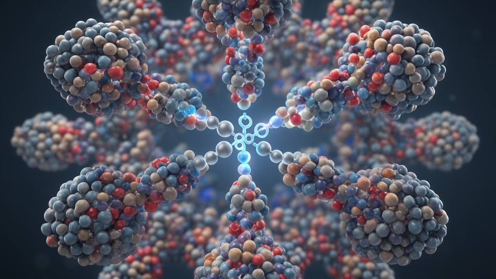
Structural modeling refined the interface to single residues. The team submitted full-length nucleoprotein and phosphoprotein sequences to the HDOCK server for global protein-protein docking without predefined binding sites, then filtered the returned models against the experimentally implicated regions. Although Model 1 achieved the most favorable docking score, it formed only one contact inside the regions of interest, whereas Model 6 formed seven clustered contacts there and was taken as the region-consistent representative conformation. Atom-level classification identified a single hydrogen-bond candidate between the glutamine 370 of the nucleoprotein and serine 254 of the phosphoprotein, with their atoms separated by 2.66 angstroms. Neighboring phosphoprotein residues Ile255, Pro256, and Met257 faced the polar glutamine and the side-chain-free glycine 371, contributing to van der Waals packing and main-chain shape complementarity rather than paired hydrophobic interactions.
Conservation analysis underscored the functional importance of this motif. Across 1,610 successfully anchored Newcastle disease virus nucleoprotein sequences retrieved from public databases, the 367 to 375 window was nearly invariant, with every position retaining its consensus residue in at least 97.64 percent of strains. The three interface residues were among the most conserved in the window: glutamine 370 was conserved in 99.81 percent of strains, alanine 369 in 99.94 percent, and glycine 371 was completely invariant across all 1,610 sequences. Subsequent co-immunoprecipitation and immunofluorescence assays confirmed that mutating either glutamine 370 of the nucleoprotein or serine 254 of the phosphoprotein significantly impaired the interaction and disrupted inclusion body-like structure formation, validating the modeled contact experimentally.
The authors argue that this contact is functionally distinct from the two canonical nucleoprotein-phosphoprotein interactions described in paramyxoviruses: the N-terminal region of the phosphoprotein that chaperones unassembled nucleoprotein to prevent premature oligomerization, and the C-terminal X domain that binds the nucleoprotein tail of the assembled template to position the polymerase. Because the multimerization domain mediates phosphoprotein oligomerization into tetramers, its engagement by the nucleoprotein provides multivalency, with each oligomer presenting multiple binding surfaces and polymeric nucleoprotein arrays presenting repeated motifs. Such multivalent, weak-affinity interactions are precisely the biophysical drivers of liquid-liquid phase separation, offering a mechanistic explanation for how a short linear motif within the nucleoprotein core can nucleate phase separation in concert with the oligomeric phosphoprotein scaffold. The researchers note that the structures of the nucleoprotein-phosphoprotein complex, and even of the nucleoprotein-phosphoprotein-polymerase assembly, still need to be resolved by cryo-electron microscopy or X-ray crystallography. They also observe that the virus’s robust replication in chicken cells reflects the natural susceptibility of avian hosts, while its oncolytic property, the selective infection and lytic destruction of cancer cells, adds translational relevance to understanding how viral condensates form.
Subject of Research: Residue Gln370 of Newcastle disease virus nucleoprotein mediate viral inclusion body-like structure formation via interaction with residue Ser254 of viral phosphoprotein
Article Title: Residue Gln370 of Newcastle disease virus nucleoprotein mediate viral inclusion body-like structure formation via interaction with residue Ser254 of viral phosphoprotein
Article References: Wang, W., Xiao, S., Zhang, Y., Yuan, X., Dong, W., Qin, Z., Hu, F., Yu, K., Barysavets, D., Ihar, N., Natallia, S., Wang, H., & Li, G. (2026). Residue Gln370 of Newcastle disease virus nucleoprotein mediate viral inclusion body-like structure formation via interaction with residue Ser254 of viral phosphoprotein. Frontiers in Cellular and Infection Microbiology, 16, Article 1978310. https://doi.org/10.3389/fcimb.2026.1978310
Image Credits: AI Generated
DOI: 10.3389/fcimb.2026.1978310
Keywords: Residue, Gln370, Newcastle, disease, virus, nucleoprotein, mediate, viral, inclusion, body-like, structure, formation
News Source: Kristina Jarvis. (October 24, 2026). Single Amino Acid Contact Drives Assembly of Newcastle Disease Virus Replication Factories. Scienmag.