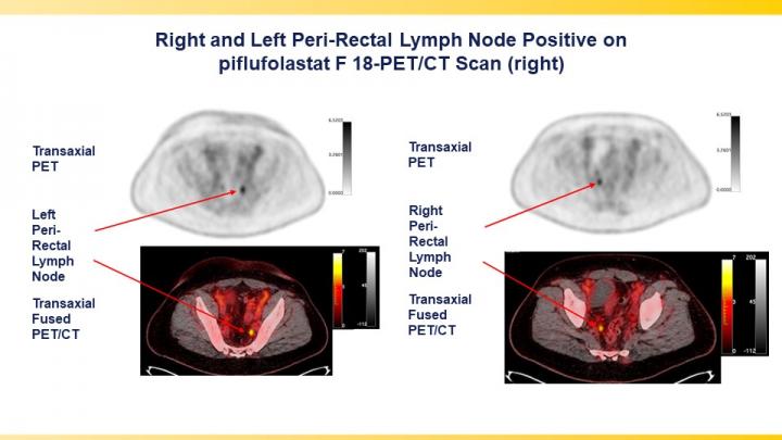
Newly FDA-approved imaging agent now available for patients

Credit: Images courtesy of Lantheus Holdings, Inc., Billerica, MA.
Reston, VA (Embargoed until 3:00 p.m. EDT, Tuesday, June 15, 2021)–A phase III clinical trial has validated the effectiveness of the prostate-specific membrane antigen (PSMA)-targeted radiotracer 18F-DCFPyL in detecting and localizing recurrent prostate cancer. Approved by the U.S. Food and Drug Administration last month, the radiotracer identified metastatic lesions with high positive predictive values regardless of anatomic region, adding to the evidence that PSMA-targeted radiotracers are the most sensitive and accurate agents for imaging prostate cancer. This study was presented at the Society of Nuclear Medicine and Molecular Imaging (SNMMI) 2021 Annual Meeting.
Prostate cancer patients have high levels of PSMA expression, which makes PSMA an effective target for imaging the disease. In previous studies, the novel positron emission tomography (PET) imaging agent 18F-DCFPyL was found to bind selectively with high affinity to PSMA. To demonstrate the diagnostic performance of 18F-DCFPyL for regulatory approval, a prospective, multicenter study was conducted in 14 sites across the United States and Canada.
The study sought to determine the positive predictive value (the probability that patients with a positive screening test actually have the disease) and detection rate of 18F-DCFPyL PET/computed tomography (CT) by anatomic region, specifically the prostate/prostate bed, pelvic lymph nodes, and regions outside the pelvis. Study participants included men who had rising prostate-specific antigen (PSA) levels after local therapy as well as negative or equivocal conventional imaging results.
Patients were imaged with 18F-DCFPyL PET/CT, then imaged again after 60 days to verify suspected lesions using a composite “standard of truth,” which consisted of histopathology, correlative imaging findings and PSA response. Comparing findings between the 18F-DCFPyL imaging and the “standard of truth,” the positive predictive value and detection rate were measured.
18F-DCFPyL-PET/CT was found to successfully detect and pinpoint metastatic lesions with high positive predictive value, regardless of their location in the body, in men with biochemically recurrent prostate cancer who had negative or equivocal baseline imaging. Higher positive predictive values were observed in extra-pelvic lymph nodes and bone compared to soft tissue regions.
With the recent approval of 18F-DCFPyL (now referred to as piflufolastat F-18) by the FDA, the impact of this research may be realized in the very near future. As these agents become more widely available, patients with newly diagnosed, recurrent, and metastatic prostate cancer may have new therapeutic approaches available to them. The results of the study will be presented at the SNMMI meeting by Steven Rowe, MD, PhD, associate professor of radiology and radiological science at Johns Hopkins University in Baltimore, Maryland.
Abstract 123. “A Phase 3 study of 18F-DCFPyL-PET/CT in Patients with Biochemically Recurrent Prostate Cancer (CONDOR): An Analysis of Disease Detection Rate and Positive Predictive Value (PPV) by Anatomic Region,” Steven Rowe and Michael Gorin, Johns Hopkins, Baltimore, Maryland; Lawrence Saperstein, Yale School of Medicine, New Haven, Connecticut; Frederic Pouliot, Departement de Chirurgie, Division d’Urologie, University of Quebec, Quebec, Canada; David Josephson, Tower Urology, Cedars Sinai Medical Center, Los Angeles, California; Peter Carroll, UCSF, San Francisco, California; Jeffrey Wong, City of Hope, Sierra Madre, California; Austin Pantel, University of Pennsylvania Health System, Philadelphia, Pennsylvania; Morand Piert, University of Michigan, Ann Arbor, Michigan; Kenneth Gage, Diagnostic Imaging and Interventional Radiology, H. Lee Moffitt Cancer Center and Research Institute, Tampa, Florida; Steve Cho, University of Wisconsin-Madison, Madison, Wisconsin; Andrei Iagaru, Stanford University, Stanford, California; Janet Pollard, University of Iowa Hospital, Iowa City, Iowa; Vivien Wong, Jessica Jensen and Nancy Stambler, Progenics Pharmaceuticals, Inc., New York, New York; Michael Morris, Memorial Sloan-Kettering Cancer Center, New York, New York; and Barry Siegel, Washington University School of Medicine, St. Louis, Missouri.
###
All 2021 SNMMI Annual Meeting abstracts can be found online at https:/
About the Society of Nuclear Medicine and Molecular Imaging
The Society of Nuclear Medicine and Molecular Imaging (SNMMI) is an international scientific and medical organization dedicated to advancing nuclear medicine and molecular imaging, vital elements of precision medicine that allow diagnosis and treatment to be tailored to individual patients in order to achieve the best possible outcomes.
SNMMI’s members set the standard for molecular imaging and nuclear medicine practice by creating guidelines, sharing information through journals and meetings and leading advocacy on key issues that affect molecular imaging and therapy research and practice. For more information, visit http://www.
Media Contact
Rebecca Maxey
[email protected]


