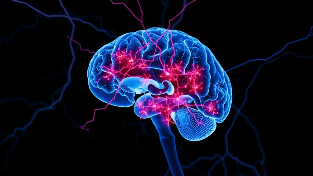
Prion diseases are among the rarest and most uniformly fatal neurodegenerative disorders known to medicine. Conditions such as Creutzfeldt-Jakob disease, fatal familial insomnia, kuru, scrapie, and bovine spongiform encephalopathy share a common pathological hallmark: the conversion of the normal cellular prion protein, PrP^C, into a misfolded, protease-resistant form, PrP^Sc. This conformational transformation, which rearranges a primarily alpha-helical structure into a beta-pleated sheet, triggers an exponential auto-catalytic accumulation of aggregates that spread between neurons and progressively destroy brain tissue. A new review published in Frontiers in Cell Death now integrates current mechanistic insights into how this proteopathic process drives neurodegeneration through the collapse of protein homeostasis, or proteostasis, and the sustained activation of the unfolded protein response, a cellular stress signaling network that can shift from adaptive protection to lethal execution.
The review, authored by Amna Syeda, Mehwish Nafiz, and Shah Ubaid-ullah, examines how misfolded prion protein perturbs endoplasmic reticulum homeostasis and thereby imposes proteotoxic stress on neurons. The endoplasmic reticulum is the major site of protein folding in the cell, assisted by a complex family of molecular chaperones. When misfolded or aggregation-prone proteins exceed the capacity of these chaperones, the ubiquitin-proteasome system, and organelle quality-control mechanisms, the accumulation of aberrant proteins becomes self-reinforcing. Aggregates sequester chaperones and quality-control factors, impair clearance pathways, disrupt organelle function, and increase the burden imposed on the ER. Neurons are particularly vulnerable to this cascade because they are long-lived, highly polarized, metabolically demanding, and unable to dilute damaged proteins through cell division.
Central to the review’s argument is the role of the unfolded protein response as a double-edged sword. Under normal conditions, the ER luminal faces of three transmembrane mediators, PERK, IRE1α, and ATF6, are bound by the chaperone BiP, which holds all three in an inactive state. When misfolded proteins accumulate in the ER lumen, BiP dissociates to bind these substrates, allowing the three mediators to activate. PERK phosphorylates the translation initiation factor eIF2α, inducing translational repression to prevent further protein overload. ATF4, which escapes this repression due to upstream open reading frames in its mRNA, is selectively translated and encodes proteins involved in autophagy and protein folding. IRE1α dimerizes and activates its endoribonuclease activity, splicing XBP1 mRNA to produce a transcription factor that induces genes for protein folding and ER-associated degradation. ATF6 translocates to the Golgi apparatus, where it is cleaved by S1P and S2P endopeptidases, releasing a fragment that moves to the nucleus to induce XBP1 and ERAD genes.
While these three branches collectively aim to restore ER proteostasis during stress, sustained activation under conditions of excessive proteotoxic burden can shift signaling toward cell-death pathways. The PERK-eIF2α axis emerges as the particularly detrimental branch in prion neurodegeneration. In infected mice overexpressing wild-type PrP^C, sustained eIF2α phosphorylation by PERK leads to a sudden decrease in synaptic protein levels by nine weeks post-infection, long before overt clinical manifestations appear. Over time, the adaptive response becomes pathological when the repression of critical synaptic proteins leads to synaptic failure and neuronal demise. Post-mortem studies of brain tissue from individuals with Creutzfeldt-Jakob disease support this evidence, showing increased levels of ER stress-related proteins and pro-apoptotic stress signaling.
Beyond neurons themselves, the review highlights how chronic PERK activation in astrocytes induces a specific neurotoxic reactivity state. Reactive astrocytes show altered expression of synapse-related and extracellular matrix-related proteins, as well as stress and inflammation indicators, leading to synaptic deficits and non-cell-autonomous neuronal toxicity. Treatments using ER stressors such as thapsigargin and tunicamycin enhance PERK-eIF2α activation and the expression of neurotoxic markers in astrocytes, while PERK inhibition reduces marker expression and preserves synaptic function and viability in prion-infected mice. By comparison, the IRE1-XBP1 pathway takes on a more nuanced and potentially redundant role in prion disease, as CNS-specific deletion of XBP1 did not substantially affect prion replication, neuronal loss, stress response, or survival of prion-infected mice. Evidence for the IRE1α-ASK1-JNK signaling pathway is stronger in related neurodegenerative models than in prion disease itself, and prion-specific evidence for ATF6 remains limited.
The review further examines how UPR signaling intersects with multiple cell-death mechanisms beyond classical apoptosis. ER calcium release stimulates the calcium-activated phosphatase calcineurin in prion-infected cells, precipitating neurodegeneration, and FK506-mediated inhibition of calcineurin significantly reduces disease progression in prion models. Autophagic dysfunction has been described both experimentally and in patients, implicating autophagy-lysosome abnormalities in prion proteostasis. PrP^Sc has been found in endolysosomal compartments, and defects in autophagosome maturation, lysosomal acidification, cargo recognition, or lysosomal membrane integrity allow undegraded material to accumulate. Because lysosomal and ER functions are coupled through lipid trafficking, calcium signaling, and organelle contacts, lysosomal failure can intensify ER stress rather than remain a separate downstream event. Ferroptotic lipid peroxidation and necroptotic signaling may also contribute to neuronal demise, and these pathways can coexist within the same tissue.
On the therapeutic front, the review discusses pharmacological strategies targeting UPR signaling and stress-response pathways to mitigate neurodegeneration. Partial restoration of protein synthesis downstream of PERK has demonstrated neuroprotective effects in prion-diseased mice without causing pancreatic toxicity, particularly in studies involving ISRIB, a small molecule that restores aspects of translation downstream of eIF2α phosphorylation. Selective targeting of the kinase insert loop of PERK has shown the ability to reduce neurodegeneration while avoiding systemic toxicity in prion models. Next-generation eIF2B activators such as DNL343 have demonstrated CNS penetrance and neuroprotective potential in models of TDP-43 and ALS that are dependent upon the integrated stress response. However, the authors emphasize that none of the UPR-targeting agents has reached clinical trials for Creutzfeldt-Jakob disease, and that findings summarized in the review are preclinical unless human trials have demonstrated safety, target engagement, and clinical benefit.
The review also positions emerging experimental platforms as critical bridges between mechanistic insight and clinical translation. Human stem cell-derived cerebral organoids now allow prion infections to be modeled in three-dimensional systems that are physiologically meaningful, capturing subtype-specific disease dynamics and enabling drug screening. Organoids capture key aspects of neuronal heterogeneity and cell-cell interactions, providing deeper insight into prion propagation and associated proteostatic stress responses. Simultaneously, advances in machine learning and multi-omics integration are accelerating the identification of UPR modulators by enabling the integration of transcriptomic, proteomic, and metabolomic datasets, providing a systems-level understanding of UPR signaling dynamics. Machine learning frameworks allow prioritization of candidate targets, prediction of drug-target interactions, and stratification of patient subgroups based on molecular signatures of proteostasis imbalance.
Despite the growing preclinical promise, the authors caution that the field’s principal limitation is an uneven evidence base. Much of the mechanistic data comes from infected rodents, cultured cells, or related proteinopathies, whereas longitudinal human measurements of UPR activity in prion disease remain difficult due to biosafety constraints and post-mortem degradation. Post-mortem tissue provides important confirmation but cannot reliably establish the timing of pathway activation. The authors argue that CSF and blood assays should be developed alongside prion-seeding assays, neurofilament measurements, and clinical staging, with explicit testing of whether UPR markers add prognostic or pharmacodynamic value. Integration of UPR biomarker assays through established Creutzfeldt-Jakob disease surveillance networks would be useful in tracking disease progression and response to treatment.
Collectively, the review supports a model in which proteostasis collapse and UPR signaling form a reinforcing network rather than a linear pathway. Misfolded PrP and related aggregate-prone proteins increase the demand on chaperones, proteasomal degradation, autophagy, lysosomes, and ER folding machinery. Failure of these systems increases aggregate persistence and promotes chronic PERK-eIF2α-ATF4-CHOP signaling, while IRE1α can couple ER stress to RIDD, ASK1-JNK signaling, inflammation, and apoptosis. ER calcium loss, mitochondrial dysfunction, and caspase activation provide additional routes to neuronal injury. The strongest preclinical rationale currently favors interventions that restore translational homeostasis or improve clearance without causing systemic toxicity. The authors conclude that selective modulation of proteostasis and UPR signaling, rather than indiscriminate pathway blockade, holds significant promise for therapeutic intervention, provided that future studies identify reliable markers of UPR activation and develop strategies that selectively modulate this pathway without disrupting physiological proteostasis.
Subject of Research: Proteostasis collapse and unfolded protein response-mediated neurodegeneration in prion disease
Article Title: Proteostasis collapse and unfolded protein response-mediated neurodegeneration in prion disease
Article References: Proteostasis collapse and unfolded protein response-mediated neurodegeneration in prion disease. (n.d.). https://doi.org/10.3389/fceld.2026.1941546
Image Credits: AI Generated
DOI: 10.3389/fceld.2026.1941546
Keywords: prion disease, unfolded protein response, PERK-eIF2α, proteostasis, neurodegeneration, Creutzfeldt-Jakob disease, ER stress, Frontiers in Cell Death, collapse, unfolded, protein, response-mediated
News Source: Diana Fleming. (October 15, 2026). Proteostasis Collapse and Unfolded Protein Response-Mediated Neurodegeneration in Prion Disease. Scienmag.