In a compelling new case report published in the March 2026 issue of Oncoscience, researchers present a diagnostically perplexing manifestation of mucinous ovarian carcinoma (MOC) masquerading as a pelvic abscess in a postmenopausal woman. This rare subtype of epithelial ovarian cancer, typically presenting in younger women aged 20 to 40, accounts for only 3 to 5 percent of ovarian malignancies. However, the unusual clinical trajectory in a 73-year-old woman challenges conventional diagnostic paradigms and underscores the critical need for heightened vigilance when evaluating adnexal masses in older patients.
The patient initially presented with nonspecific but persistent symptoms, including abdominal pain and postmenopausal bleeding, which are often dismissed or attributed to benign conditions. Preliminary diagnostic imaging suggested an infectious etiology—specifically a pelvic abscess—prompting conservative management rather than immediate oncologic intervention. This initial misclassification illustrates a significant pitfall in clinical diagnostic reasoning, where overlapping radiologic features can obscure the underlying neoplastic process.
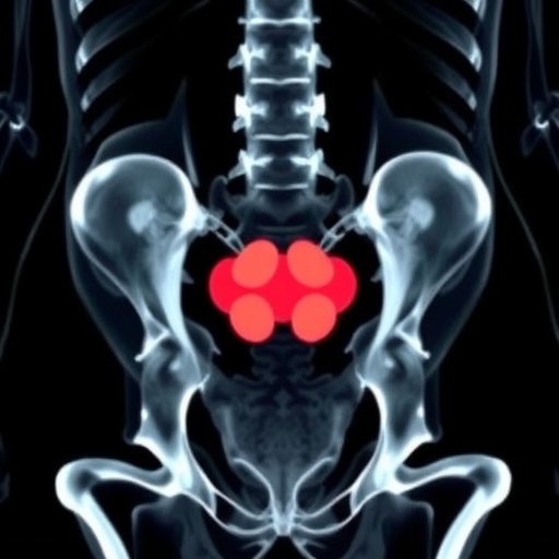
Subsequent progressive symptoms necessitated re-evaluation using advanced radiologic modalities. Magnetic resonance imaging (MRI) identified a complex multiloculated lesion with solid and cystic components in the adnexal region, diverging from typical benign infectious presentations. Concomitantly, serum tumor markers, including CA-125, carcinoembryonic antigen (CEA), and human epididymis protein 4 (HE4), were markedly elevated. These biomarkers, often leveraged in ovarian cancer diagnostics, raised a strong suspicion for malignancy despite the patient’s atypical demographic profile.
Surgical intervention ensued, with comprehensive staging and histopathological confirmation revealing stage IC2 mucinous adenocarcinoma of the ovary. Tumor histology demonstrated hallmark features of intestinal-type differentiation, characterized by glandular proliferation and villoglandular architecture infiltrating the ovarian stroma. Notably, the presence of goblet cells within the epithelium highlighted phenotypic heterogeneity uncommon in ovarian neoplasms but typical in gastrointestinal tract malignancies, further complicating the diagnostic picture.
This case accentuates the deceptive clinical course of mucinous ovarian carcinoma, particularly in postmenopausal patients, where malignancies are less anticipated. The clinical overlap between inflammatory/infectious pathologies and neoplastic processes poses profound challenges, potentially delaying definitive diagnosis and curative treatment. The findings advocate for a multidisciplinary approach, combining imaging studies, serological tumor markers, and expert pathological assessment to expedite accurate diagnosis in ambiguous adnexal masses.
Therapeutically, the patient underwent optimal cytoreductive surgery, followed by adjuvant chemotherapy with carboplatin and paclitaxel regimens. Despite the development of postoperative infectious complications, aggressive management facilitated recovery and continuation of adequate oncologic care. This favorable short-term outcome reinforces the importance of early recognition and comprehensive treatment strategies tailored to individual patient profiles.
From a pathophysiological perspective, mucinous ovarian carcinoma’s rarity and histogenetic complexity contribute to diagnostic challenges. Its intestinal-type differentiation suggests a possible origin from transitional or metaplastic epithelium, setting it apart from the more common serous subtypes. Furthermore, the disease’s propensity to present with large multiloculated cystic masses filled with mucinous material can mimic benign cystadenomas or inflammatory abscesses on imaging, highlighting the necessity for advanced diagnostic tools and clinical suspicion.
The elevation of tumor markers such as CA-125, while nonspecific, gains critical diagnostic weight when interpreted alongside CEA and HE4 levels, especially in patients with atypical presentations. These markers not only aid in the initial diagnostic workup but also serve as valuable tools for postoperative monitoring and detection of disease recurrence. Their combined assessment should be incorporated routinely when imaging results are equivocal.
This report also invites a reevaluation of clinical guidelines for postmenopausal women presenting with adnexal masses, challenging the tendency toward conservative management based solely on initial benign imaging impressions. The biological behavior of mucinous carcinoma, with its potential for aggressive local infiltration even in early stages, necessitates a more proactive diagnostic and therapeutic stance to improve long-term survival outcomes.
The multidisciplinary collaboration detailed in this case includes gynecologic oncologists, radiologists, and pathologists, reflecting the imperative for integrated care models in managing complex gynecologic malignancies. The synergy of clinical expertise with advanced technological diagnostics exemplifies precision medicine principles, aiming to tailor interventions according to nuanced disease characteristics and patient factors.
In conclusion, this exceptional case of mucinous ovarian carcinoma masquerading as a pelvic abscess in a postmenopausal woman serves as a critical reminder of the diagnostic complexities within gynecologic oncology. It stresses the importance of comprehensive evaluation protocols that transcend initial clinical impressions, advocating for the integration of imaging, serum biomarkers, and histopathology to discern malignancy in ambiguous pelvic masses. Heightened clinical suspicion coupled with multidisciplinary input can significantly alter patient outcomes, ensuring timely intervention in rare but formidable ovarian cancers.
DOI link for further details and full access to the case report is available at https://doi.org/10.18632/oncoscience.650.
Subject of Research: People
Article Title: Deceptive clinical course of mucinous ovarian carcinoma mimicking pelvic abscess in a postmenopausal woman: An exceptional case report
News Publication Date: 11-Mar-2026
Web References: https://doi.org/10.18632/oncoscience.650
Image Credits: Copyright © 2026 Jarathi et al., CC BY 4.0 License
Keywords: cancer, mucinous cystadenocarcinoma, pelvic abscess, carcinoma ovary, histopathological diagnosis
Tags: adnexal mass evaluation in elderlyclinical presentation of mucinous ovarian carcinomadelayed cancer diagnosis in postmenopausal womendiagnostic challenges in ovarian cancerelevated CA-125 and ovarian cancerepithelial ovarian cancer subtypesMRI imaging for ovarian tumorsmucinous ovarian carcinoma case reportovarian cancer misdiagnosed as pelvic abscessovarian cancer tumor markers CEA HE4pelvic abscess differential diagnosispostmenopausal ovarian cancer diagnosis

