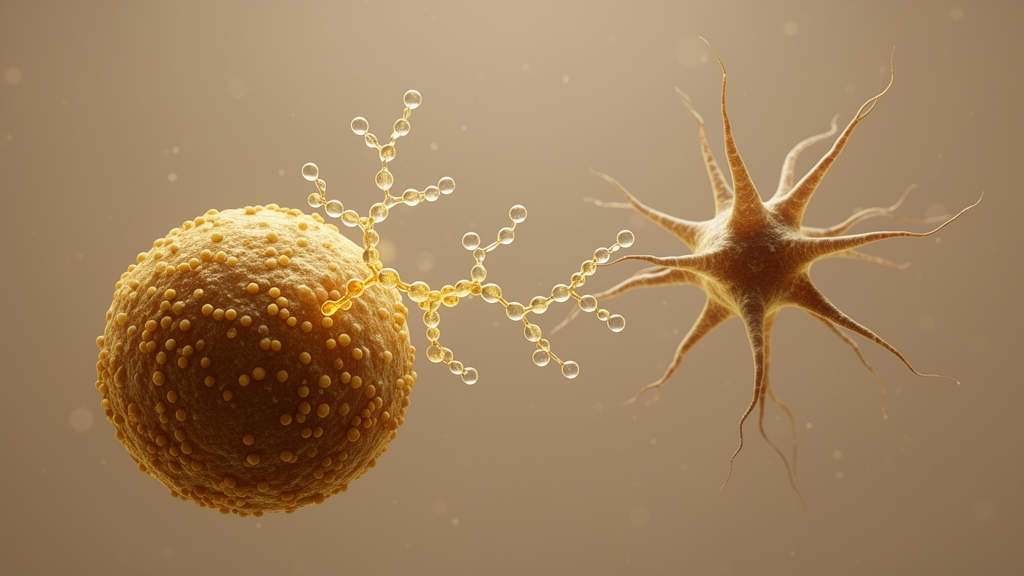
Researchers have mapped the fine structure of a complex pectic polysaccharide isolated from the pollen of Typha angustifolia, a plant long used in East Asian traditional medicine, and shown in laboratory experiments that the molecule can drive mouse dendritic cells toward a mature, antigen-presenting state. The polysaccharide, designated PTPS-1-3, carries an unusual combination of structural domains, including arabinogalactan-II-rich side chains, homogalacturonan segments, and diagnostic molecular signatures associated with rhamnogalacturonan-II, one of the most intricate and highly conserved motifs found in plant pectins. When the team exposed bone marrow-derived dendritic cells from mice to the purified polysaccharide, the cells up-regulated key co-stimulatory molecules, secreted more inflammatory cytokines, and became better at activating antigen-specific CD8-positive T cells in a co-culture system. The work, published in the journal Chinese Medicine, pairs an unusually detailed structural assignment with functional immune testing, addressing a long-standing obstacle in polysaccharide research: the sheer structural heterogeneity of pectins has made it difficult to pin down which molecular features actually carry biological activity.
The structural challenge is central to the field. Unlike small drug-like molecules with defined architectures, plant polysaccharides are heterogeneous, hierarchically organized chains whose biological behavior depends on chain size, monosaccharide composition, linkage types, branching patterns, and the presence of uronic acid-rich domains. Pectins are especially complicated, built from smooth homogalacturonan regions interspersed with hairy regions rich in rhamnogalacturonan-I and rhamnogalacturonan-II. Classical tools such as methylation analysis and nuclear magnetic resonance spectroscopy can struggle with overlapping signals and low-abundance motifs, and arabinogalactan-II-rich side chains and rhamnogalacturonan-II-related structures are notoriously hard to resolve within intact pectic matrices. The new study set out to overcome that limitation for PTPS-1-3, a fraction previously isolated in the group’s earlier work from Typha angustifolia pollen, which had already hinted at arabinogalactan-II and rhamnogalacturonan-II features but whose detailed organization and immunological properties remained largely unexplored.
The isolation began with commercially obtained pollen authenticated as Typha angustifolia L., drawn from a single batch, and followed the team’s previously established extraction and purification protocol. Size-exclusion analysis by high-performance gel permeation chromatography showed a single, symmetric peak corresponding to a molecular weight of approximately 10 kilodaltons, placing PTPS-1-3 firmly in the low-molecular-weight range for a pectic polysaccharide. Earlier colorimetric testing based on the thiobarbituric acid reaction had suggested rhamnogalacturonan-II-associated structural signatures, motivating the deeper structural investigation. Complementary physical characterization reinforced the pectic identity: infrared spectroscopy revealed a broad O-H stretching absorption, carboxylate bands, a strong glycosidic vibration, and a peak indicating esterified or protonated carboxyl groups consistent with uronic acid-containing domains, while scanning electron microscopy showed irregular sheet-like aggregates with a loose, porous surface architecture that the authors note is consistent with amorphous aggregation of highly branched polysaccharide chains after drying.
Chemical analysis established the sugar backbone of the molecule. PTPS-1-3 contained roughly 19.2 percent uronic acid, confirming its classification as an acidic pectic polysaccharide, and its monosaccharide profile was dominated by rhamnose, arabinose, xylose, galactose, and galacturonic acid. The relatively high arabinose and galactose content pointed to arabinogalactan-type domains, while rhamnose and galacturonic acid indicated rhamnogalacturonan backbone structures, and the detection of xylose together with the earlier thiobarbituric acid-positive result supported possible rhamnogalacturonan-II-related motifs. To sharpen the picture, the researchers prepared two derivatives: a reduced form in which uronic acid residues were converted to allow linkage detection, and a partial acid hydrolysate designed to strip away acid-labile side chains and enrich acid-resistant structural segments. Methylation analysis of the intact polysaccharide identified nineteen partially methylated alditol acetate derivatives, a sign of high branching and structural heterogeneity, with major linkages including terminal and 1,5-linked and 1,3,5-linked arabinofuranose and terminal, 1,3-linked, 1,6-linked, and 1,3,6-linked galactopyranose patterns characteristic of arabinan and arabinogalactan side chains.
The reduction step proved particularly informative. After uronic acid reduction, new 1,4-linked and 1,3,4-linked galactopyranose residues appeared at relative abundances of 10.7 percent and 10.6 percent respectively, residues likely derived from reduced galacturonic acid units and supporting the presence of homogalacturonan segments and substituted galacturonic-acid-containing regions. The partial acid hydrolysate, by contrast, showed markedly reduced proportions of the arabinofuranosyl linkages and an increased proportion of terminal galactopyranose, indicating that arabinofuranosyl residues sit mainly in acid-labile side chains attached to galactan-rich structures. The authors caution that several rare monosaccharide residues characteristic of rhamnogalacturonan-II domains normally occur at low abundance and are difficult to fully identify by gas chromatography-mass spectrometry-based methylation analysis alone, so complete assignment of the rhamnogalacturonan-II-related features required the multidimensional NMR work that followed.
That spectroscopic work delivered the study’s most detailed structural evidence. Recording proton, carbon, DEPT135, COSY, HSQC, and HMBC spectra on a 600 MHz instrument, the team assigned proton and carbon signals to twenty-nine distinct glycosyl residues spanning arabinose, galactose, xylose, galacturonic acid, rhamnose, fucose, arabinopyranose, glucuronic acid, and apiose units. Downfield anomeric carbon signals at 108 to 110 parts per million marked arabinose, while signals at 103 to 106 parts per million corresponded to galactose, and the coexistence of abundant beta-galactopyranose backbones substituted with alpha-arabinofuranosyl side chains emerged as a diagnostic feature of arabinogalactan-II. Signals characteristic of galacturonic acid confirmed the homogalacturonan domain, with non-esterified and esterified 1,4-linked alpha-galacturonic acid units assigned at distinct chemical shifts. Crucially, the proton spectrum contained signals assigned to H-3 protons of Kdo and Dha, two hallmark components of rhamnogalacturonan-II side chains that are difficult to detect by monosaccharide composition analysis alone, along with methyl group signals from rhamnose residues in four substitution patterns, a fucose signal, and the rare 1,3-linked beta-apiofuranosyl residue, whose attachment chemistry matches the conserved rhamnogalacturonan-II architecture in which complex side chains attach to O-2 of galacturonic acid residues. Long-range HMBC correlations established transglycosidic connectivities supporting thirty-two assigned linkages. The authors are careful to note that the arabinogalactan-II and homogalacturonan assignments were directly supported by their own compositional, linkage, and NMR data, whereas the rhamnogalacturonan-II-related signals were identified by comparison with previously reported spectra, and a tentative structural model was proposed on that combined basis.
With the structure in hand, the functional testing turned to dendritic cells, the specialized antigen-presenting cells that bridge innate and adaptive immunity. Bone marrow-derived dendritic cells generated from eight-week-old C57BL/6 mice were treated with PTPS-1-3 at concentrations from 5 to 40 micrograms per milliliter for 24 hours. A CCK-8 viability assay showed that treatment significantly increased cell viability, reaching approximately 150 to 190 percent of the control level, with viability remaining above control at all tested concentrations, indicating no detectable cytotoxicity within that range. Quantitative PCR and ELISA measurements showed significantly up-regulated mRNA expression and markedly increased secretion of the pro-inflammatory cytokines TNF-alpha, IL-1 beta, and IL-6. Flow cytometry revealed markedly increased surface expression of the co-stimulatory molecules CD40, CD80, and CD86 on CD11c-positive cells, while FITC-dextran uptake assays showed significantly reduced phagocytic capacity. Together, these changes trace the characteristic transition of immature dendritic cells, which excel at antigen capture but express few co-stimulatory molecules, into mature antigen-presenting cells primed to launch adaptive responses.
Transcriptomic analysis added a genome-wide dimension. RNA sequencing of treated and control dendritic cells, analyzed with differential expression thresholds of a q value below 0.05 and fold change greater than 2 or below 0.5, revealed substantial transcriptional alterations, and KEGG pathway enrichment showed that the differentially expressed genes concentrated in immune-related pathways including antigen processing and presentation, cytokine-cytokine receptor interaction, cell adhesion molecules, and T cell activation-related pathways. Gene set enrichment analysis independently confirmed significant enrichment of the T cell activation involved in immune response gene set. The functional consequence was tested in an OT-I co-culture system, in which dendritic cells pretreated with PTPS-1-3, ovalbumin, both, or neither were co-cultured with purified CD8-positive T cells from OT-I mice, whose T cells recognize a specific ovalbumin peptide. PTPS-1-3 combined with ovalbumin significantly promoted T cell proliferation measured by Ki67 staining and increased the proportions of IFN-gamma-positive and TNF-alpha-positive CD8-positive T cells compared with ovalbumin alone, whereas PTPS-1-3 alone did not substantially activate the T cells in the absence of antigen. That pattern suggests the polysaccharide mainly enhances dendritic cell-mediated antigen presentation rather than directly provoking non-specific T cell activation.
A preliminary structure-activity comparison rounded out the study. Intact PTPS-1-3 induced the strongest up-regulation of CD40, CD80, and CD86 among the tested polysaccharides, outperforming both its own partial acid hydrolysate, which lost arabinose-rich side chains, and FPLP, a previously characterized homogalacturonan-type polysaccharide from Ficus pumila fruit whose galacturonic acid-rich backbone resembles the homogalacturonan regions of PTPS-1-3 but lacks the complex arabinogalactan-II-rich side chains and rhamnogalacturonan-II-related features. FPLP showed relatively weak activity, suggesting that a homogalacturonan-like backbone alone is insufficient to account for the full maturing effect, while the weakened activity of the hydrolysate points to a contribution from the intact, branched domain organization. The authors are explicit about the caveats: partial acid hydrolysis also reduces molecular weight, which is known to influence polysaccharide-receptor interactions, so the observed differences may reflect combined effects of structural alteration and molecular size, and studies using polysaccharides of comparable molecular weight will be needed to clarify the precise relationship. Further limitations include the unknown receptor-binding sites and the need for receptor-blocking strategies, pathway inhibitors, receptor-deficient models, and in vivo validation.
Subject of Research: Structural characterization and dendritic cell-modulating activity of the pectic polysaccharide PTPS-1-3 from Typha angustifolia pollen
Article Title: Structural characterization of an RG-II-associated complex pectic polysaccharide from Typha angustifolia pollen with dendritic cell-modulating activity
Article References: Xu, Y., Guo, M., Liu, Y., Chen, J., Shi, S., Shi, C., Wang, S., & Wang, H. (2026). Structural characterization of an RG-II-associated complex pectic polysaccharide from Typha angustifolia pollen with dendritic cell-modulating activity. Chinese Medicine, 21(1), Article 224. https://doi.org/10.1186/s13020-026-01515-9
Image Credits: AI Generated
DOI: 10.1186/s13020-026-01515-9
Keywords: Typha angustifolia, pectic polysaccharide, rhamnogalacturonan-II, dendritic cells, BMDC maturation, CD8+ T cells, antigen presentation, NMR spectroscopy, methylation analysis, immunomodulation, structure-activity relationship, OT-I co-culture
News Source: Ophelia Keating. (October 29, 2026). Pollen Polysaccharide With Complex Pectic Architecture Boosts Dendritic Cell Maturation. Scienmag.