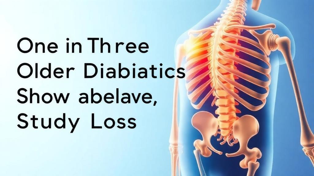
More than one in three older adults living with type 2 diabetes may be harboring dangerously weakened bones without knowing it, according to a new cross-sectional study from Iran that adds fresh urgency to a long-underappreciated complication of the world’s fastest-growing chronic disease. Researchers at Tabriz University of Medical Sciences found that 32.4 percent of patients with type 2 diabetes mellitus aged over 54 years met the diagnostic threshold for osteoporosis, a figure that rivals prevalence rates seen in much older general populations and that far exceeds what most clinicians would expect in a cohort whose average age barely crossed the mid-fifties.
The study, published in BMC Endocrine Disorders, recruited 102 patients from the endocrinology clinics and wards of Imam Reza and Sina Hospitals in Tabriz. Each participant underwent dual-energy X-ray absorptiometry, the gold-standard imaging technique for measuring bone mineral density, with readings taken at the lumbar spine and the femoral neck, the upper portion of the thigh bone where most hip fractures originate. Following World Health Organization criteria, osteoporosis was defined as a T-score of -2.5 or lower at either site. When degenerative changes in the spine, common in older adults, threatened to distort lumbar measurements, the researchers gave priority to the femoral neck values, a methodological safeguard designed to avoid falsely inflating or deflating the true burden of disease.
The resulting prevalence estimate, 32.4 percent with a 95 percent confidence interval of 23.4 to 42.3 percent, carries a wide margin of uncertainty, reflecting the modest sample size. Even at the lower bound, however, the numbers suggest that nearly a quarter of older diabetic patients in this setting have bones fragile enough to fracture under everyday stresses. That matters because diabetes-related fractures are not ordinary fractures. Studies across multiple populations have shown that people with type 2 diabetes can break hips and vertebrae at bone mineral density levels that would be considered safe in non-diabetic individuals, meaning standard screening thresholds may systematically underestimate their true skeletal risk.
The biological explanation lies partly in a process that has fascinated bone biologists for years: the accumulation of advanced glycation end products, or AGEs. Chronic high blood sugar drives glucose molecules to bind irreversibly to proteins throughout the body, including collagen, the structural scaffold of bone. Glycated collagen becomes stiffer and more brittle, and the normal microscopic repair process, in which bone cells remove and replace damaged tissue, slows down. The result is a bone that may look adequately dense on a scan but behaves mechanically like aged plastic rather than resilient composite material. Diabetes also disrupts the delicate coupling between osteoblasts, the cells that build bone, and osteoclasts, the cells that dismantle it, tilting the balance toward loss.
Against this backdrop, the Tabriz team used logistic regression to identify which characteristics of their patients were actually associated with osteoporosis. Two factors emerged clearly and independently. The first was female sex: women in the cohort had roughly five times the odds of osteoporosis compared with men, an unadjusted odds ratio of 4.84 with a confidence interval of 1.67 to 14.01 and a P value of 0.004. After adjusting for other variables, the association persisted, with an adjusted odds ratio of 3.34 and a P value of 0.035. The finding aligns with decades of bone biology, since the sharp decline in estrogen at menopause removes a powerful protective signal that normally restrains bone resorption, and it suggests that postmenopausal women with diabetes face a compounding of two skeletal threats at once.
The second independent predictor was more surprising to many observers: kidney function. The researchers measured the estimated glomerular filtration rate, or eGFR, a standard laboratory index of how efficiently the kidneys filter blood, expressed in milliliters per minute per 1.73 square meters of body surface area. Lower eGFR was significantly associated with osteoporosis, with an odds ratio of 0.96 per unit increase, a confidence interval of 0.94 to 0.98, and a P value of 0.004. In the multivariable model the relationship held, with an adjusted odds ratio of 0.97 and a P value of 0.037. In practical terms, each incremental decline in filtration capacity nudged patients measurably closer to the osteoporotic range.
The kidney-bone connection is well grounded in physiology. The kidneys are not passive bystanders in skeletal metabolism; they activate vitamin D, regulate calcium and phosphate balance, and orchestrate the hormonal signals that keep bone remodeling on schedule. As filtration declines, phosphate retention and hormonal disturbances trigger a cascade known as chronic kidney disease-mineral and bone disorder, in which parathyroid hormone levels rise and bone turnover becomes disordered. Because diabetic kidney disease is itself one of the most common complications of long-standing type 2 diabetes, the study’s finding effectively identifies a subgroup, older diabetic women with waning renal function, whose bones are being attacked from multiple directions simultaneously.
What the study did not find is equally instructive. Glycemic control, measured through fasting blood sugar and glycated hemoglobin, body mass index, physical activity levels assessed with the International Physical Activity Questionnaire, and medication histories did not emerge as statistically significant independent predictors in this cohort. That may reflect the limited statistical power of a 102-patient sample, or genuine homogeneity among patients recruited from specialized endocrinology services, where most participants receive broadly similar care. The authors are careful on this point, and their conclusion emphasizes that the two robust signals, female sex and reduced eGFR, should guide screening priorities rather than serving as an exhaustive risk map.
The clinical implications are straightforward but consequential. Current guidance in many health systems does not routinely recommend bone density screening for patients with type 2 diabetes until they reach ages well beyond the range of this cohort, partly because diabetes modestly raises body weight, which is traditionally protective against bone loss, and partly because the fracture risk of diabetic patients was underappreciated for years. This study, conducted in a real-world hospital population in northwestern Iran, argues that waiting is a mistake. Integrating a simple DEXA scan and a kidney function check into the routine management of diabetic patients over 55, with heightened vigilance for women, could identify high-risk individuals years before a first fragility fracture, which is often the moment the disease announces itself.
The researchers, led by Yalda Taati Alamdari and corresponding author Mahsa Malekian, with support from the Research Vice-Chancellor of Tabriz University of Medical Sciences, acknowledge the limits of a single-center, cross-sectional design, which captures a snapshot in time and cannot establish whether low kidney function causes bone loss or merely travels alongside it. They call for prospective multicenter studies to sharpen risk stratification and inform screening strategies. With more than half a billion people worldwide now living with diabetes, and populations aging fastest in exactly the regions where diabetes is expanding most rapidly, the study’s central message travels far beyond Tabriz: in older patients with type 2 diabetes, the skeleton is a silent organ of the disease, and the tools to catch its decline already sit in every endocrinology clinic.
Subject of Research: Prevalence and risk factors of osteoporosis among older patients with type 2 diabetes mellitus
Article Title: Prevalence and associated factors of osteoporosis in type 2 diabetes mellitus patients: a cross-sectional study
Article References: Taati Alamdari, Y., Lotfi Yagin, N., Gilani, N., Sadra, V., Najafipour, F., Gaffarzadeh Rad, S., & Malekian, M. (2026). Prevalence and associated factors of osteoporosis in type 2 diabetes mellitus patients: a cross-sectional study. BMC Endocrine Disorders. https://doi.org/10.1186/s12902-026-02525-2
Image Credits: AI Generated
DOI: 10.1186/s12902-026-02525-2
Keywords: type 2 diabetes, osteoporosis, bone mineral density, DEXA, eGFR, kidney function, postmenopausal women, fragility fractures, advanced glycation end products, cross-sectional study, Iran, endocrinology
News Source: Ophelia Keating. (October 14, 2026). One in Three Older Diabetics Show Silent Bone Loss, Study Finds. Scienmag.