Extracellular vesicles circulating in the blood of young mice do not rescue injured kidney tubule cells, and neither do vesicles from old mice, according to a laboratory study published in BMC Molecular and Cell Biology. Instead, the decisive factor turned out to be the age of the macrophages themselves: conditioned medium from bone marrow-derived macrophages taken from old mice protected hypoxia-stressed tubular epithelial cells more effectively than medium from young macrophages, regardless of any vesicle pre-treatment. The finding runs counter to a popular narrative in which young extracellular vesicles rejuvenate aged tissues and old vesicles accelerate decline.
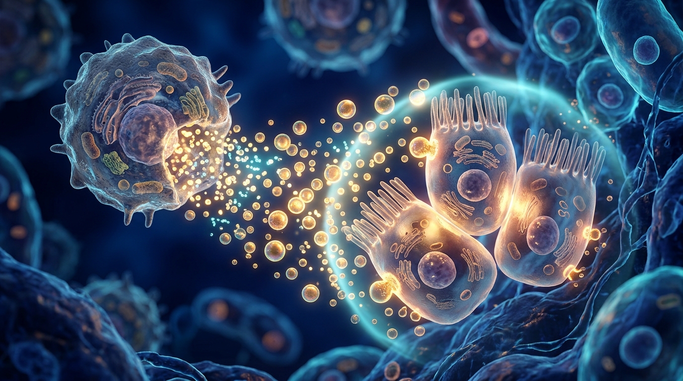
Acute kidney injury is a major global health problem that raises the risk of death and of progression to chronic kidney disease, and supportive care remains the mainstay of treatment because effective therapies to limit renal injury are limited. Hypoxic injury to tubular epithelial cells triggers inflammation, apoptosis, necrosis and epithelial-mesenchymal transition, a process in which epithelial cells take on mesenchymal characteristics and can contribute to later fibrosis. Pro-inflammatory mediators such as tumor necrosis factor-alpha, interleukin-6 and the chemokine CCL2 are released during this response, alongside established injury biomarkers including kidney injury molecule 1 and lipocalin-2. Macrophages sit at the centre of this crosstalk: early in injury their secretions can amplify tubular damage, while during recovery reparative macrophage secretomes promote tubular cell survival and resolution of inflammation.
The research team, led by Dora Livkisa and David J. Lundy with colleagues including Chia-Te Liao, set out to test whether the age of circulating plasma vesicles changes how macrophages respond to stressed kidney cells. They used extracellular vesicles previously isolated from the plasma of young male C57BL/6 mice aged 7 to 12 weeks and aged male mice aged 70 to 90 weeks, following International Society for Extracellular Vesicles guidelines. The same vesicle batches had been characterised in an earlier study, which found that vesicles from old mice were slightly smaller, at 93.0 versus 116.6 nanometres, and present at lower concentrations, and that a 752-microRNA panel revealed only a limited set of differences in cargo between the age groups.
Because the hypoxia system relied on sealed anaerobic boxes that could not expose only one cell type to low oxygen, the researchers adopted a sequential design rather than a simultaneous co-culture. Tubular epithelial cells were first subjected to hypoxia followed by reoxygenation, using a 30-hour hypoxia and 6-hour reoxygenation protocol. Bone marrow-derived macrophages were then stimulated with conditioned medium from hypoxic or normoxic tubular cells, with or without prior priming with young or old vesicles, and the resulting macrophage-conditioned media were finally applied to fresh injured tubular cells. The team measured cell viability with CCK-8 assays and quantified gene expression by RT-qPCR, using Hnrnpab as the reference gene.
The direct test was unambiguous. Untreated hypoxia-reoxygenation injury reduced tubular cell viability to 56.3 percent of normoxic levels. Adding young vesicles left viability at 50.1 percent and old vesicles at 45.8 percent, neither significantly different from the untreated injured group, and the two vesicle types did not differ from each other. Plasma vesicles therefore showed no acute direct protective effect on hypoxia-injured tubular epithelial cells under these conditions.
The indirect route through macrophages told a more layered story. Naive macrophages from young mice responded to hypoxic tubular cell secretome with increased Il1b and Il10 expression, whereas macrophages from old mice showed no significant gene expression changes, indicating that macrophages from older mice were less transcriptionally responsive to the injury stimulus. When the macrophages were first primed with vesicles, their responses to the hypoxic secretome grew far larger. Young macrophages primed with young vesicles showed decreased Il6 and Nos2 and increased Arg1 and Il10, a broadly anti-inflammatory pattern, while priming with old vesicles produced a mixed response including increased Arg2 and Ccl17 and a small decrease in Il10. Old macrophages primed with young vesicles showed decreased Il1b and Il6 and greatly increased Arg1, whereas old vesicle priming increased Il6, Tnfa, Arg1 and Ccl17. A clustering analysis of all macrophage gene expression data showed a significant response in M2-associated genes when macrophages of either age were primed with old vesicles, but not with young vesicles.
When the researchers applied macrophage-conditioned medium to hypoxia-injured tubular cells, the results overturned their expectations. Untreated injury dropped viability to 54.3 percent, and conditioned medium from naive macrophages improved survival above non-conditioned medium, showing that the macrophage secretome contains protective compounds even without deliberate polarisation. But medium from naive old-donor macrophages protected significantly better, at 78.4 percent viability, than medium from naive young-donor macrophages, at 67.4 percent. Pre-treatments modified protection within each age group, with lipopolysaccharide-stimulated young macrophages more protective than interleukin-4-stimulated ones, yet vesicle priming made no significant difference: young macrophages treated with young or old vesicles yielded viabilities of 66.2 and 63.6 percent against 67.4 percent for naive cells, and old macrophages treated with young or old vesicles yielded 80.3 and 79.3 percent against 78.4 percent for naive cells. A two-way analysis of variance across the vesicle-primed conditions confirmed a significant main effect of macrophage donor age on viability, with no main effect of vesicle donor age and no interaction between the two factors.
Inflammatory and fibrotic gene expression in primary tubular epithelial cells followed the same pattern. Hypoxia drove clear increases in Il6 and Ccl2, and conditioned medium from macrophages polarised with lipopolysaccharide produced the highest Il6 expression. In the vesicle-primed groups, two-way analysis showed significant main effects of macrophage donor age on Il6 and Ccl2 expression, while vesicle donor age was not significant for either transcript and no interactions were detected. Epithelial-mesenchymal transition markers behaved similarly: hypoxic injury alone triggered a robust EMT response, with Fn1 and Acta2 showing the largest inductions and the epithelial marker Cdh1 falling, while under normoxia all markers stayed at baseline. The young macrophage secretome had little effect on EMT marker expression regardless of pre-treatment, whereas the old macrophage secretome was significantly less stimulative of Acta2 expression than the young secretome. Again, macrophage donor age showed significant main effects on Fn1 and Acta2, vesicle donor age did not, and no interactions were found.
The authors are careful about what these results do and do not show. They note that macrophages differentiated ex vivo from young and aged mice may differ not only in chronological age but also in progenitor composition and baseline epigenetic or metabolic states, so the observed effects may reflect age-associated reprogramming during differentiation rather than ageing of mature macrophages. They also acknowledge that the sequential tubular cell-macrophage-tubular cell design sacrifices the realism of a living kidney, that viability assays and transcriptional signatures in vitro may indicate early anti-inflammatory and anti-fibrotic effects that do not necessarily translate into full cytoprotection or reduced fibrosis in vivo, and that donor-age differences in macrophage metabolism could have altered the composition of the conditioned media. The team used both HK-2 cells and primary tubular epithelial cells and found the main qualitative trends consistent across both models, and they deliberately used vesicles from healthy donors rather than septic or ischaemic animals, since such stimuli profoundly remodel vesicle cargo. Macrophage activation states were inferred from M1- and M2-associated genes without protein-level measurements, a limitation the authors flag alongside the absence of proteomic or metabolomic analyses.
What remains striking is the reversal of the expected age gradient. Prior work by the same group had shown that young plasma vesicles consistently attenuated pro-inflammatory gene expression in macrophages stimulated with lipopolysaccharide, and other studies have reported that young plasma vesicles improved mitochondrial metabolism and functional performance across multiple organs in aged mice. In this disease-relevant context, however, oldness was neither inherently pro-inflammatory nor pro-fibrotic: old macrophage secretomes suppressed tubular inflammation, did not exaggerate EMT responses and rescued viability more effectively than young secretomes. The authors suggest that the modest cargo differences between young and old vesicles, profiled across 752 microRNAs, may explain why vesicle age did not dramatically alter tubular cell outcomes, and that vesicle cargo-driven effects are context-dependent, varying with isolation methods, characterisation and dosing. The study was conducted under approval from the Taipei Medical University Institutional Animal Care and Use Committee and used non-SPF-housed mice, and the authors position the work as hypothesis-generating, pointing toward future tests of vesicles from injured donors and in vivo models to determine how age and acute injury together shape vesicle function in kidney repair.
Subject of Research: How macrophage age and circulating extracellular vesicles affect protection of hypoxia-injured kidney tubular cells
Article Title: Investigating the roles of macrophage age and circulating extracellular vesicles in cytoprotection of hypoxic renal tubular cell stress
Article References: Livkisa, D., Lundy, D. J., Lee, T.-L., Szomolay, B., & Liao, C.-T. (2026). Investigating the roles of macrophage age and circulating extracellular vesicles in cytoprotection of hypoxic renal tubular cell stress. BMC Molecular and Cell Biology, 27(1), Article 24. https://doi.org/10.1186/s12860-026-00583-x
Image Credits: AI Generated
DOI: 10.1186/s12860-026-00583-x
Keywords: acute kidney injury, extracellular vesicles, macrophages, hypoxia, tubular epithelial cells, ageing, cytokines, epithelial-mesenchymal transition, bone marrow-derived macrophages, inflammation, kidney repair, cell culture
News Source: Drew Townsend. (November 4, 2026). Old Macrophage Secretomes Outperform Young Ones in Protecting Stressed Kidney Cells. Scienmag.