Hypertrophic obstructive cardiomyopathy, long managed with blunt tools such as beta blockers and open-heart surgery, is entering a new pharmacological era. A comprehensive review published in Frontiers in Cardiovascular Medicine on 27 October 2026 by Chukwunenyem Nwuba of Mid and South Essex NHS Foundation Trust and Jason N. Dungu of Anglia Ruskin University maps the full treatment landscape for the condition, from decades-old drugs to the cardiac myosin inhibitors mavacamten and aficamten, and asks how these targeted agents might reorder a treatment pathway that has remained largely unchanged for generations.
Hypertrophic cardiomyopathy is defined as increased left ventricular wall thickness or mass exceeding 15 millimetres that cannot be explained by loading conditions such as hypertension. In children, the wall thickness must exceed two standard deviations above the predicted mean for body surface area. The disease is typically inherited in an autosomal dominant fashion, driven by variants in sarcomere protein genes, with MYH7, MYBPC3, TNNT2 and TPM1 together accounting for roughly 30 to 40 percent of cases. Historically estimated to affect about one in 500 people, newer estimates suggest prevalence could be as high as one in 300 or one in 200, making it the most common identifiable cause of sudden cardiac death in young people under 35. Yet overall survival for most patients is comparable to an age- and sex-matched population without heart disease, and many carriers never develop clinically detectable disease, with reported penetrance of 57 percent.
At the molecular level, the disease disrupts the energetic economy of the heart muscle. In a healthy heart, 40 to 50 percent of myosin heads sit in an energy-sparing off state; in hypertrophic cardiomyopathy, this proportion is cut by more than half, leaving only 15 to 20 percent switched off. The surplus active myosin heads consume extra adenosine triphosphate and bind actin more readily, generating excessive cross-bridges during both contraction and relaxation. The result is inefficient, overly forceful contraction and impaired filling, which over time triggers pro-hypertrophic, pro-inflammatory and pro-fibrotic pathways, producing fibrosis, myocyte disarray and increased mechanical stress. When this hypertrophy narrows the outflow tract — through thickening of the interventricular septum and its interaction with the mitral valve’s systolic anterior motion — the condition is classified as the obstructive form, which is associated with dangerous ventricular arrhythmias.
Traditional drug therapy remains the first step. Non-vasodilating beta blockers, titrated to the maximum tolerated dose, have been recommended since the mid twentieth century; they reduce the outflow tract gradient by slowing heart rate and contractility, improving diastolic filling. Evidence comparing individual agents is sparse, but one of the few randomised trials showed metoprolol significantly reduced outflow gradients before, during and after exercise by 47, 34 and 70 millimetres of mercury respectively, with improved symptoms and quality of life. In the BASIC study, bisoprolol produced a mean gradient reduction of 28 millimetres of mercury, with 36 percent of patients falling below 30 millimetres and 33 percent improving by at least one New York Heart Association class. The non-dihydropyridine calcium channel blockers verapamil and diltiazem offer alternatives, though verapamil is contraindicated with beta blockers because of the risk of severe bradycardia, hypotension and conduction block, and should not be used in severe obstruction or raised pulmonary pressure because of the risk of pulmonary oedema.
For patients who remain symptomatic, disopyramide — a class IA antiarrhythmic with strong negative inotropic effects — is added on top of an atrioventricular nodal blocking agent rather than used alone. It is particularly effective at lowering outflow gradients in adults, but carries anticholinergic side effects including constipation, urinary retention, blurred vision and dry mouth, requires routine QTc monitoring with dose reduction above 500 milliseconds, and is contraindicated with other QT-prolonging drugs, glaucoma and prostatism. A cautionary note comes from a 2023 study of 51 children with the condition, the largest to date in childhood use, in which early symptomatic improvement exceeding 80 percent was not maintained in 33 percent of patients — a tachyphylaxis phenomenon that may limit the drug’s long-term value as a way of deferring escalation. Supply chain problems in the United Kingdom have further complicated its use.
When drugs fail, invasive septal reduction therapy takes over. Surgical ventricular septal myectomy, the primary surgical option, is an open-heart procedure performed under general anaesthesia with cardiopulmonary bypass, in which hypertrophied basal septal tissue is excised through an aortotomy under transoesophageal echocardiographic guidance. It reduces outflow tract gradients in more than 90 percent of patients, and concomitant procedures — mitral valve repair in about 9 percent of cases, coronary bypass grafting in roughly 5 percent, and Cox-Maze IV ablation for atrial fibrillation — address associated anomalies in around 30 percent. A 2005 study of 289 patients reported survival of 98, 96 and 83 percent at one, five and ten years, comparable to the general population, while a study following 1,999 children over more than 45 years found operative mortality of just 1 percent, with redo myectomy needed in 5.8 percent at five years and 8.3 percent at ten years.
Alcohol septal ablation offers a minimally invasive alternative: one to three millilitres of 96 to 100 percent ethanol is injected into a targeted septal branch of the left anterior descending coronary artery, inducing a controlled localised infarction that thins the septum and widens the outflow tract over weeks to months. It works best in patients with septal thickness of at least 17 millimetres and favourable coronary anatomy, and about 90 percent of patients improve to New York Heart Association class I or II. But it carries a pacemaker requirement in 5 to 15 percent of cases, ventricular arrhythmias in 1 to 2 percent, and a risk of coronary damage. Generally, myectomy is preferred for younger, lower-risk patients with marked hypertrophy above 30 millimetres or complex mitral and subvalvular anatomy, while ablation suits older or higher-risk patients unsuitable for surgery. A 2023 study of 19,007 patients treated between 2010 and 2019 found surgical outcomes depended strongly on hospital volume, whereas ablation outcomes did not — though low-volume operators were associated with increased 30-day mortality for both procedures.
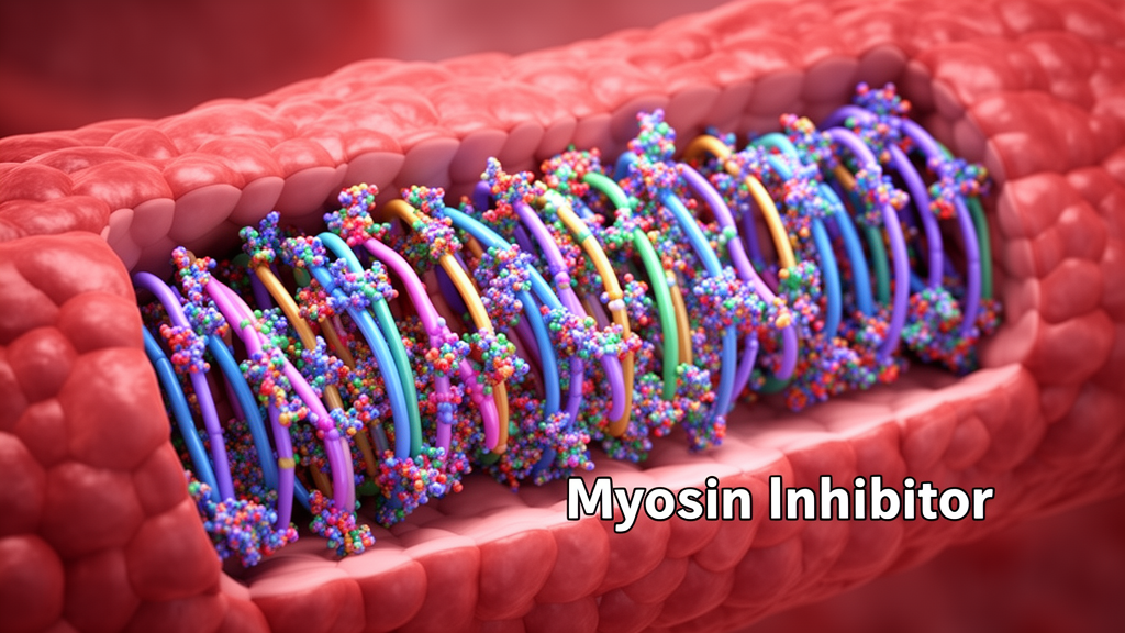
Into this landscape come the myosin inhibitors, which directly address the disease’s core mechanism by stabilising myosin heads in their inactive, ATP-bound state, limiting cross-bridge cycling and reducing contractility. Mavacamten, approved by the FDA in 2022 and by the MHRA and NICE in 2023, was the first in class. In the pivotal EXPLORER-HCM trial — a phase 3, randomised, double-blind, placebo-controlled study of 251 adults with New York Heart Association class II or III symptoms and outflow gradients of at least 50 millimetres of mercury despite background therapy — 37 percent of mavacamten patients met the primary composite functional endpoint versus 17 percent on placebo, with significant gradient reduction, symptom improvement and generally good tolerability, though a small number experienced reversible ejection fraction reductions requiring monitoring. Benefits have been sustained through 180 weeks of extension follow-up, with up to 80 percent of patients achieving elimination of obstruction after six months in subsequent studies.
Aficamten, a second-in-class agent approved by the FDA and NICE, is building its own evidence base rapidly. The SEQUOIA-HCM phase 3 trial of 282 participants showed improved exercise capacity, symptoms, outflow gradients and mitral regurgitation, with serious adverse events in only 5.6 percent; among patients eligible for septal reduction at baseline, 88 percent were no longer eligible after 24 weeks. The MAPLE-HCM study demonstrated aficamten’s superiority over metoprolol in obstructive disease, raising the possibility that myosin inhibitors could challenge beta blockers as default first-line therapy in selected patients. Similarly, the VALOR-HCM trial found that only 17.9 percent of mavacamten-treated patients referred for septal reduction remained eligible at 16 weeks, compared with 76.8 percent on placebo. These findings suggest many patients once destined for surgery may be managed pharmacologically instead.
Important limits remain. The drugs act slowly, typically needing four to eight weeks for significant gradient reduction, and achieve reductions of 30 to 50 millimetres of mercury — substantial, but less consistently complete than myectomy’s near-universal reductions below 10 to 20 millimetres. Discontinuation leads to recurrence of obstruction, and lifelong echocardiographic surveillance of ejection fraction is required to avoid excessive systolic suppression. In non-obstructive disease the picture is mixed: mavacamten failed to improve exercise capacity or health status over placebo in the 580-person ODYSSEY-HCM trial, while aficamten met its primary endpoint in ACACIA-HCM, though its effect on peak oxygen uptake (0.64 millilitres per kilogram per minute) was far smaller than in the obstructive SEQUOIA trial (1.8). Longer-term durability data, cost-effectiveness analyses, access and monitoring burden remain unresolved, and the authors note that treatment choice — including gene therapy, which has so far shown promise only in mouse models — should be individualised through a multidisciplinary team balancing anatomy, comorbidities and patient preference.
Subject of Research: Treatment options for hypertrophic obstructive cardiomyopathy, including traditional drug therapies, septal reduction procedures and the emerging role of cardiac myosin inhibitors
Article Title: Treatment options for hypertrophic obstructive cardiomyopathy in the era of myosin inhibitors
Article References: Nwuba, C., & Dungu, J. N. (2026). Treatment options for hypertrophic obstructive cardiomyopathy in the era of myosin inhibitors. Frontiers in Cardiovascular Medicine, 13, 1814860. https://doi.org/10.3389/fcvm.2026.1814860
Image Credits: AI Generated
DOI: 10.3389/fcvm.2026.1814860
Keywords: hypertrophic obstructive cardiomyopathy, mavacamten, aficamten, myosin inhibitors, septal myectomy, alcohol septal ablation, left ventricular outflow tract obstruction, beta blockers, disopyramide, EXPLORER-HCM, SEQUOIA-HCM, sudden cardiac death
News Source: Ophelia Keating. (October 27, 2026). New myosin inhibitors reshape treatment of obstructive hypertrophic cardiomyopathy. Scienmag.