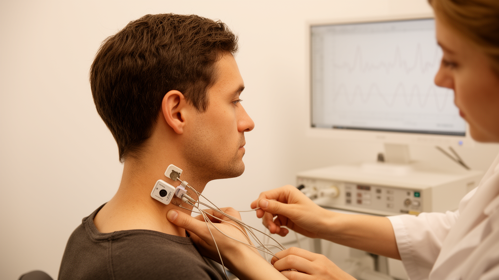
A noninvasive electrical stimulation technique applied to the neck can shift markers of parasympathetic nervous system activity and brain rhythms in healthy adults, but only when the stimulation settings are tuned to each individual, according to a pilot study published in Bioelectronic Medicine. Across four laboratory sessions, twenty healthy volunteers received stimulation at varying frequencies, current intensities, electrode placements, and durations while the team recorded heart rate variability and electroencephalography. No single universal setting stood out. Instead, each participant showed a distinct favored frequency, and selecting that personalized frequency produced substantially larger responses than any one fixed frequency applied to the whole group.
The study, registered with ClinicalTrials.gov as NCT04100486, was designed around two biomarkers. The first was the root mean square of successive R-R interval differences, or RMSSD, a heart rate variability measure that reflects beat-to-beat variation driven by vagal efferent activity to the heart. The second was global EEG alpha-band power in the 8 to 13 Hz range, which prior work has associated with parasympathetic-dominant states and high vagal tone. Participants, eleven men and nine women with a mean age of 31.2 years, attended four morning sessions separated by at least a week. Each visit tested one stimulation parameter at a time. Every condition was delivered in a twelve-minute block consisting of four minutes of baseline recording, four minutes of stimulation, and four minutes of post-stimulation recording, with conditions randomized within each session.
In the first visit, four frequencies were compared: 10, 25, 40, and 150 Hz. Current intensity was raised as high as each participant could tolerate without pain or discomfort, up to a ceiling of 400 microamps, with electrodes in a bilateral neck configuration. The result was striking in its absence of a group-level winner: no single frequency produced a statistically significant increase in RMSSD or alpha-band power across the cohort. Yet when the researchers looked at individuals, every participant had a ranked preference. Selecting each person’s favored frequency yielded a mean RMSSD increase of 41 percent, compared with a mean change of only 4.6 percent for any single frequency applied to all subjects. A null simulation, repeated 10,000 times, confirmed that this personalization gain exceeded what would be expected from simply picking the maximum of four random measurements, with a p value of 0.0003.
The neural side of the data told a parallel story. Alpha-band power also showed a preferred frequency for each participant, producing an average increase of 24 percent, compared with a maximum of 11 percent from 40 Hz stimulation and less for the other frequencies. Personalization based on the cortical marker was somewhat less effective than personalization based on RMSSD, but the pattern was consistent: the same stimulation protocol could push one person’s vagal and cortical markers up while leaving another’s flat, depending on frequency. The favored frequency appeared at random positions within the testing visit rather than clustering at the beginning or end, which argues against simple order effects driving the individual preferences.
The second visit probed current intensity. Each participant received only the frequency that had produced their largest RMSSD response in visit one, now tested at three levels: half of the sensation threshold, the sensation threshold itself, and a high intensity that remained tolerable and comfortable. High-intensity stimulation dominated. In twelve of twenty participants, or 60 percent, the supra-sensation level produced the largest RMSSD response, compared with three participants at sensation threshold and five at half threshold. Alpha-band power showed a similar tilt, with the high intensity winning in half of the participants. Notably, 60 percent of participants tolerated stimulation at the full 400-microamp ceiling. The authors note that sub-threshold intensities, which would be more comfortable in clinical settings and easier on device batteries, produced much weaker responses in this healthy cohort.
The third visit varied electrode montage: bilateral neck electrodes with contralateral torso returns, left-side-only stimulation, or right-side-only stimulation, all at high intensity and each person’s favored frequency. Here the findings ran against standard clinical practice. Left-sided stimulation is the historical default for implanted vagus nerve stimulation, largely to avoid cardiac complications, since the right vagus nerve passes near the sinoatrial node and could theoretically provoke bradyarrhythmias. In this study, however, left-only stimulation decreased RMSSD relative to baseline, with a mean decrease of 19 percent, while right-sided stimulation increased it by 9 percent on average and bilateral stimulation by 14 percent. Right-sided placement produced the largest RMSSD response in 55 percent of participants. For alpha-band power, both bilateral and right-sided montages increased the signal, winning in 35 and 45 percent of participants respectively, while left-only stimulation left the average unchanged.
A complication emerged across visits two and three: when frequency and other parameters were held constant, the RMSSD response faded over time, dropping from 41 percent in visit one to 31 percent and then 14 percent. The researchers interpret this as possible habituation or regression, and it motivated a recalibration step. In the fourth visit, frequency testing was repeated with high intensity and right-side-only stimulation. The recalibrated favored frequency produced a mean RMSSD increase of 54 percent, again exceeding the null expectation for selecting the maximum of four random draws, with a p value of 0.036. But the preferred frequency itself had shifted in 75 percent of participants between visits one and four, and the alpha-band personalization gain similarly rebounded from 9 percent to 39.5 percent after recalibration. The authors argue this makes intermittent re-checking of personalized settings a likely necessity in any future clinical protocol.
Duration produced the most mixed picture. In visit four, a twenty-minute stimulation block was compared against the four-minute blocks used throughout the study, using each person’s recalibrated frequency at high intensity on the right side. Normalized post-stimulation responses were generally smaller after the longer session, and alpha-band power actually dropped for the longer duration. Because ECG recordings are overwhelmed by stimulation artifact, the team added a finger photoplethysmography sensor in visit four, validating its derived pulse rate variability against simultaneous ECG with a correlation coefficient of 0.7439. This continuous measure revealed oscillatory autonomic dynamics during stimulation: the response cycled roughly every two minutes, especially in the first eight minutes, with a peak parasympathetic response around four minutes and another elevated response near eleven minutes before returning toward baseline. The authors suggest that up to four minutes of stimulation may be sufficient to promote an RMSSD increase, with longer stimulation potentially plateauing or diminishing the effect.
The team is careful about what the study does and does not show. The cohort was small, healthy, and aged 22 to 41, significant responses were not always measured, and inter-individual variability was high, with no parameter set working universally. There was no sham condition, though all comparisons were within participant and within visit, and the authors state that any future efficacy claim in a defined disease population will require sham-controlled design. They also note that, as with other noninvasive approaches targeting the neck or ear, there remains no confirmation of actual vagus nerve engagement, which is why they use the neutral term TCES rather than claiming cervical vagus nerve stimulation. Visit order was not randomized, which could have contributed to the declining response, and EEG was averaged across the whole brain rather than analyzed regionally. Still, the authors conclude that TCES can modulate cardiac vagal and cortical responses as measured by RMSSD and alpha-band power, and that a personalized, biomarker-guided approach to parameter selection will be essential before the technique can be applied to conditions involving autonomic dysfunction.
Subject of Research: Effects of varying transcutaneous cervical electrical stimulation parameters on autonomic and neural responses in healthy adults
Article Title: Autonomic and neural responses to varying transcutaneous cervical electrical stimulation parameters
Article References: Debnath, S., Fylaktou, F., Gurfein, B. T., & Zanos, T. P. (2026). Autonomic and neural responses to varying transcutaneous cervical electrical stimulation parameters. Bioelectronic Medicine, 12(1), Article 16. https://doi.org/10.1186/s42234-026-00210-2
Image Credits: AI Generated
DOI: 10.1186/s42234-026-00210-2
Keywords: transcutaneous cervical electrical stimulation, vagus nerve stimulation, heart rate variability, RMSSD, EEG alpha-band power, autonomic nervous system, parasympathetic activity, personalized neuromodulation, bioelectronic medicine, pilot study, healthy adults, stimulation parameters
News Source: Denise Maddox. (October 28, 2026). Neck Electrical Stimulation Shows Personalized Effects on Vagal Activity in Pilot Study. Scienmag.